Continuing experiences with RIS and PACS
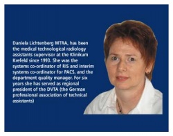
Daniela Lichtenberg MTRA
A short story about life with and without PACS - Part II - written at the Klinikum Krefeld, Germany and the Kantonssptial, Liestal, Switzerland.
In our April issue, radiographer Helga Fischer described experiences with a new PACS at the University Hospital for Radiodiagnostics, Allgemeines Krankenhaus, Vienna, Austria (EH issue 2/05. P.17. ‘PACS: The never-ending story’). In response, Daniela Lichtenberg, medical technological radiologist assistant (MTRA) at Klinikium Krefeld, Germany and F. Thanhofer*, at
the Kantonsspital Liestal, Switzerland, describe PACS experiences at their hospitals.
The 1,100-bed Klinikum Krefeld, is a maximum care institution with 21 clinical departments. In 1995 ours was one of the first hospitals to go digital and, frankly, at that point I had no clue about what a fully digital hospital would be like - or how it would work.
Here I will mention a few basic facts that need to be considered when analogue technology is being digitised:
• To avoid fear and rumours the entire staff should be informed about restructuring from Day 1
• Each step requires careful planning - that goes without saying. The more staff is involved the better the new equipment and structures will be accepted. Particularly older colleagues tend to be scared of so much new technology. When they are given sufficient and adequate information early on or - even better - can visit a department where digitisation has already been implemented, they will understand that everyone can learn.
When we started with the introduction of a RIS/PACS our department was really outmoded and thus was entirely refurbished. To install the cabling for the new system, ceilings were opened. Walls were torn down or added. We received not one CT and one angio, but an entire department equipped by Siemens. In addition to 5 bucky workstations and 3 X-ray workstations, we also received one thorax workstation. MRI was the only modality that was not replaced.
Parallel with this refurbishment, the Medos RIS was installed by a team of four. Over a period of four month the RIS was customised to reflect the entire range of services provided by the department. All screens and menus for the different user groups were configured. Since we had to do this in addition to our regular workload, our days were often very long. As soon as this phase was completed the staff was trained. A six-week test phase followed immediately, then the RIS was integrated into our routine operations - this all went without a hitch. The RIS contained digital dictaphones from the very beginning, which meant that the typing pool could immediately access dictated files. Speech recognition was added during a system upgrade in 2004.
The PACS, also by Siemens, was implemented step-by-step. Diagnostic work stations in our department are MagicViews 1002 with two screens each. When we first introduced the system we had to think a lot about light and space. Today, with flat screens, space is no longer a big issue.
Since the RIS and PACS are by two different manufacturers there is no synchronised access to patient data, which means the patient record has to be accessed both in the RIS and PACS. We solved this problem by introducing barcode scanners.
In the course of time the wards received MagicViews 200 which only allow viewing and do not have diagnostic features. Today, all wards have web-based systems.
It wasn’t easy to focus on the new technology in the middle of all the noise and dirt. But when, once again, one of us accidentally printed 60 single films because the layout function hadn’t been set to 12 images per film, we all knew: We are not going to miss the old system.
However, we made one major mistake: We introduced a RIS and PACS before, one year later, the entire hospital followed suit with a KIS. We are still struggling with a few problems due to that decision:
• The conventions of the RIS and PACS didn’t correspond to those of the KIS
• Patient data of one whole year had to be entered in the PACS
• The unique patient-ID (PAT-ID) of the KIS has not been completely integrated into the RIS and the PACS (both have their own ID administration)
• KIS overrules RIS and the data must be adjusted manually in the PACS.
We all know that today’s technology is short-lived, nevertheless it was a bit of a shock when after only eight years we had to face the fact that once we had been digital pioneers but now we were already old-fashioned.
In the meantime we had increased our short-term archive from 720 Gbyte to 2,56 Tbyte and adjusted it to the CT and MRI data volumes and flow. After eighteen months, we already had off-line MODs shelved. Processing the requested MODs became extremely time-consuming. We consequently outsourced long-term archiving. Today, this task is being taken care of by ISIS/Telepaxx.
Since digital mammography is a stand-alone unit within the hospital, the images by Sectra, which we have had for about 18 months, are archived separately.
The advantages and disadvantages mentioned by Helga Fischer in her article in European Hospital perfectly reflect my own experience. From the very beginning we had a systems co-ordinator: a MTRA from our own department was trained as systems co-ordinator. In our department he is the contact for all PACS-related issues and he is in close contact with Siemens. That means problems can be detected early on and don’t necessarily lead to down times. The biggest advantage for the MTRA was the fact that the number of mis-exposures was reduced to almost nil.
The biggest disadvantages resulted from the fact that we had tried to save money in the wrong place since we did not understand the necessity of certain investments. For example, we had to wait for three years for a post-processing station for the MTRAs, because it had not been included in the initial plan and afterwards there was no money left to purchase it.
Last but not least I should mention that the road to the digital department is not only hilly but can also be rather thorny. We consistently refused to print images for departments that had a MagicView station and thus were able to view the digital images. With that policy we did not necessarily move up in the ‘nice department’ ranking and many a ward physician developed very innovative bribery schemes to get hold of hardcopies after all. But to no avail - thanks to the fact that our boss showed backbone and to the administration’s decision to simply no longer order films.
Finally, ten points that summarise the major issues:
1. Plan every step and integrate the staff
2. Have a KIS with the necessary interface in place before you go ahead with RIS and PACS.
3. Look for fast image transfer in the network
4. Make sure the RIS and PACS are implemented immediately after the staff has been trained
5. Test!
6. Make sure the naming conventions fit for KIS, RIS and PACS
7. Ensure system maintenance from Day 1
8. Convert without compromise
9. Develop a downtime concept
10. Do follow-up training.
Today, all imaging modalities, including ultrasound and post-processing in the operating theatre (OT) are linked to the PACS and have a DICOM work list. To remain up-to-date and implement future software updates in the PACS we now have to modernise our hardware. All diagnostic workstations will be equipped with faster computers and more storage capacity (RAM and ROM). The operating system will change from Unix to Windows, which will allow synchronised access of patient records in the RIS and PACS.
Very often it turned out that the planning was good, but somehow didn’t work in reality. Therefore, it is very important to be open, creative and flexible. There are always new things to look at and to learn and new challenges to be tackled - that can only be accomplished with the co-operation of all hospital staff and staff in the companies concerned. Boredom is definitely a thing of the past. Every change should be an improvement, or, as our boss would say:
There are no problems.
There are only solutions!
07.08.2006