

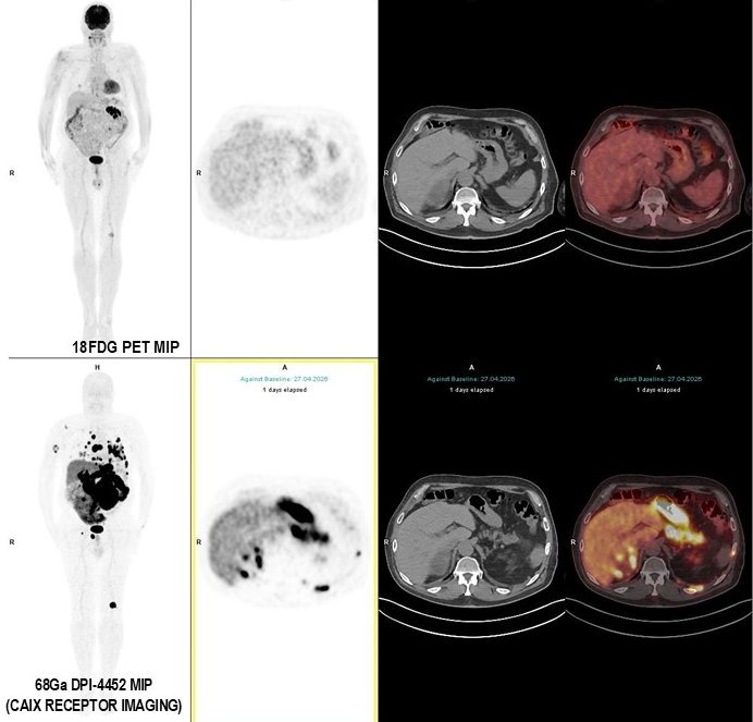
Case: In a patient diagnosed with metastatic clear cell RCC, foci observed at very low levels on 18F-FDG PET/CT, and some that were not detectable at all, were seen as dramatically dense on [68Ga]Ga-DPI-4452-PET/CT (carbonic anhydrase IX receptor) imaging.
Image courtesy of Prof. Arslan
News • Nuclear medicine
ccRCC kidney cancer: PET/CT imaging approach improves lesion detection
Research was presented at the 39th Annual Congress of the European Association of Nuclear Medicine (EANM’26)
This is according to research presented at the 39th Annual Congress of the European Association of Nuclear Medicine (EANM’26).1
Renal cell carcinoma (RCC) is the most common form of kidney cancer, with ccRCC its most common subtype.2 While 18F-FDG is commonly used as a radiotracer in cancer PET/CT imaging, it has limitations in RCC, driving interest in more targeted imaging approaches.3-7
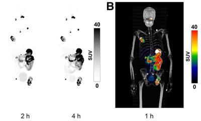
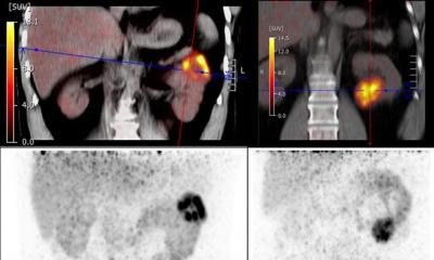
Against this background, researchers investigated a different imaging approach targeting carbonic anhydrase IX (CAIX), a protein highly expressed in ccRCC but with minimal expression in normal renal tissue. In a prospective multicentre study of 68 patients with RCC, researchers compared the CAIX-targeted imaging agent [68Ga]Ga-DPI-4452 PET/CT with 18F-FDG PET/CT.3

Photo courtesy of Prof. Arslan
Across 492 true lesions evaluated in the study, [68Ga]Ga-DPI-4452 PET/CT detected 90.9%, compared with 56.7% using 18F-FDG PET/CT. The difference was even greater in ccRCC, where [68Ga]Ga-DPI-4452 PET/CT detected 97.9% of lesions compared with 47.1% using 18F-FDG PET/CT. Patient-level diagnostic accuracy in ccRCC was also higher, at 98.2% compared with 77.2%.
“CAIX gives us a highly selective biological target,” explained lead author Professor Esra Arslan. “It is expressed at a very high rate in ccRCC, while expression in normal renal tissue remains minimal. This gives us the potential to distinguish tumour from surrounding healthy tissue much more clearly.”
By contrast, 18F-FDG measures glucose metabolism, but its effectiveness in RCC can be limited by high physiological glucose uptake and intense urinary excretion in the kidneys, which can make tumours harder to distinguish from surrounding tissue. Some low-grade tumours also take up relatively little 18F-FDG, making them more difficult to detect.
Tumour uptake with [68Ga]Ga-DPI-4452 was approximately five times higher than with 18F-FDG, while the tumour-to-background ratio – a measure of how clearly a tumour stands out from surrounding tissue – was around ten times greater. Higher lesion detection was seen across several metastatic sites, including the lymph nodes, lungs, bones, brain and soft tissues.
The difference was particularly striking in brain metastases, where high physiological 18F-FDG uptake can make lesions difficult to distinguish. [68Ga]Ga-DPI-4452 PET/CT detected all identified brain metastases, with a median maximum tumour-to-background ratio of 281.3 compared with 0.7 for 18F-FDG. [68Ga]Ga-DPI-4452 PET/CT also detected all identified lung metastases compared with around half using 18F-FDG PET/CT, while lymph-node detection was 95.0% versus 68.9%.
One exception was liver metastases. Across the overall RCC cohort, FDG performed better in the liver, reflecting lower and more variable CAIX expression in some non-clear cell RCC subtypes. Within the ccRCC subgroup, however, performance again favoured [68Ga]Ga-DPI-4452.
Importantly, [68Ga]Ga-DPI-4452 PET/CT resulted in a change in clinical management or disease stage in 30.9% of patients, compared with 5.9% using 18F-FDG PET/CT. Changes included identifying previously undetected recurrent or residual disease, detecting nodal or distant metastases that resulted in upstaging and reclassifying some patients from oligometastatic to more widespread metastatic disease. In three patients, findings considered positive on 18F-FDG PET/CT were excluded by [68Ga]Ga-DPI-4452 PET/CT, which the researchers reported avoided unnecessary interventions.
Looking ahead, the researchers, Professor Arslan and Professor Dr Halil Kömek, who contributed equally to this work, see potential roles for CAIX-targeted PET/CT in initial staging before nephrectomy or metastasectomy, restaging when recurrence is suspected and, in the future, as part of a theranostic approach in which [68Ga]Ga-DPI-4452 PET/CT could help identify patients suitable for treatment with [177Lu]Lu-DPI-4452.
“[68Ga]Ga-DPI-4452 has the potential to become a valuable tool across several stages of RCC management,” said Professor Arslan. “However, it is premature to claim that this agent represents a ‘new molecular imaging standard’ without validation in larger, multicentre studies stratified by subtype.”
Further research will be needed to validate the findings in larger patient populations and across different RCC subtypes, where CAIX expression can be lower and more variable. Longer-term prospective studies will also be important to determine whether the observed changes in disease staging and clinical management ultimately translate into improved patient outcomes.
References:
- Arslan E, Kömek H, Can C, Akgün E, Çermik TF, Kılıç R, et al. Beyond FDG: CAIX-targeted PET achieves near-complete tumor detection in renal cell carcinoma. Presented at: 39th Annual Congress of the European Association of Nuclear Medicine (EANM’26); 20 October 2026; Vienna, Austria. Presentation OP-624.
- Cleveland Clinic. Renal cell carcinoma. Available at: https://my.clevelandclinic.org/health/diseases/24906-renal-cell-carcinoma. Accessed 21 September 2026.
- Kömek H, Arslan E, Can C, Akgün E, Kılıç R, Yücetaş U, et al. From metabolism to molecular phenotype: carbonic anhydrase IX-targeted [68Ga]Ga-DPI-4452 PET/CT in renal cell carcinoma: a prospective multicenter comparison with [18F]FDG PET/CT. J Nucl Med. 2026 Sep 17:jnumed.126.273011. doi:10.2967/jnumed.126.273011. [Epub ahead of print]
- Arslan E, Kömek H, Can C, Akgün E, Türk O, Yücetaş U, et al. One tracer, whole syndrome: CAIX-targeted PET unmasks the multisystem tumor landscape of von Hippel-Lindau disease. Clin Nucl Med. 2026 Sep 24. doi:10.1097/RLU.0000000000006674. [Epub ahead of print]
- Boellaard R, Delgado-Bolton R, Oyen WJG, Giammarile F, Tatsch K, Eschner W, et al. FDG PET/CT: EANM procedure guidelines for tumour imaging: version 2.0. Eur J Nucl Med Mol Imaging. 2015;42(2):328–354. doi: 10.1007/s00259-014-2961-x.
- Franquet E, Park H. Molecular imaging in oncology: Common PET/CT radiopharmaceuticals and applications. Eur J Radiol Open. 2022;9:100455. doi:10.1016/j.ejro.2022.100455.
- Bex A, Abu Ghanem Y, Albiges L, Bonn S, Campi R, Capitanio U, et al. European Association of Urology Guidelines on Renal Cell Carcinoma: The 2025 Update. Eur Urol. 2025;87(6):683–696. doi: 10.1016/j.eururo.2025.02.020.
Source: European Association of Nuclear Medicine
20.10.2026