Image source: adapted from: Ikeo S, Tani Y, Sawayama J et al., Biomaterials 2026 (CC BY-NC 4.0)
News • Dynamic in vitro platform
“Breathing” organoid offers new insights into lung disease
Researchers at the Institute of Industrial Science, The University of Tokyo have developed a system that allows human lung organoids to expand in a breathing-like manner by applying pressure from inside the tissue.
The platform enables quantitative measurement of lung compliance—a mechanical indicator of how easily the lung expands—and may provide a new way to study diseases such as pulmonary fibrosis.
The researchers present their findings in the journal Biomaterials.
The lungs repeatedly expand and contract with each breath, and this mechanical flexibility is essential for normal gas exchange. However, most experimental lung models developed to date cannot reproduce these dynamic mechanical behaviors. Conventional organoid cultures replicate aspects of three-dimensional tissue structure but remain static systems without controlled mechanical stimulation.
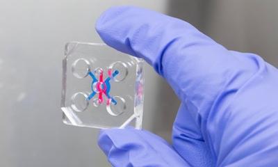
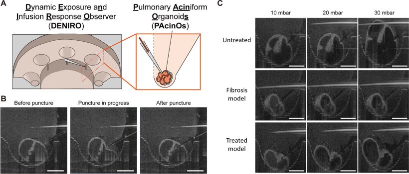
To address this limitation, the research team developed Pulmonary Aciniform Organoids (PAcinOs) derived from human induced pluripotent stem cells. These organoids mimic the structure of the pulmonary acinus, the peripheral region of the lung where gas exchange occurs.
By giving the organoids this dynamic behavior, we believe it will become possible to study lung function and disease states in conditions that more closely resemble those in the human body
Shoji Takeuchi
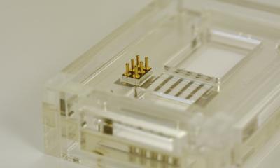
The researchers also designed a custom culture device called the Dynamic Exposure and Infusion Response Observer (DENIRO). This device allows researchers to access the internal lumen of organoids and apply controlled pressure.
By combining the PAcinO with the DENIRO platform and inserting a fine glass capillary into the organoid, its structure can be monitored using optical coherence tomography. This setup allows researchers to gradually increase the pressure inside the organoid and record the resulting three-dimensional volume changes.
Using this system, the researchers quantified lung compliance in lung organoids by measuring the relationship between pressure and volume changes.
When organoids were treated with bleomycin, a compound widely used to induce pulmonary fibrosis in experimental models, the organoids showed reduced expansion in response to pressure, indicating decreased compliance. Treatment with the antifibrotic drug nintedanib partially restored this response.
These findings demonstrate that mechanical changes associated with lung disease can be evaluated in a human induced pluripotent stem cell-derived three-dimensional organoid system.

Image source: Institute of Industrial Science, The University of Tokyo
Researcher Shoji Takeuchi said: "The lung is an organ that naturally expands and contracts with each breath. While conventional organoids have been able to reproduce lung morphology, they have not been able to reproduce its movement. We felt that a lung model that does not move is somehow incomplete, so we attempted to introduce motion by applying pressure from inside the organoid. I still remember the moment when the organoids expanded beautifully, the entire lab cheered. By giving the organoids this dynamic behavior, we believe it will become possible to study lung function and disease states in conditions that more closely resemble those in the human body. In the future, beyond applications in medicine and drug discovery, we hope this technology could also contribute to new systems such as respiratory components for biohybrid robots."
By enabling simultaneous observation of organoid structure and mechanical function, the platform provides a new experimental approach for studying lung diseases characterized by tissue stiffening.
The researchers suggest that the system may support studies of pulmonary fibrosis and other respiratory diseases, and aid the evaluation of potential therapeutic drugs in human organoid models.
Source: Institute of Industrial Science, The University of Tokyo
01.04.2026