Image source: CNIC
News • CVD prevention
3D matrix ultrasound to identify cardiovascular injury
A new imaging technique for real 3D vascular ultrasound could become a key tool in strategies aimed at preventing cardiovascular disease in apparently healthy persons, complementing traditional risk parameters such as cholesterol and high blood pressure.
The new results, published in JACC: Cardiovascular Imaging, show that real 3D vascular ultrasound is reliable, accurate, and faster than previous methods for the assessment of plaque volume in the carotid and femoral arteries.
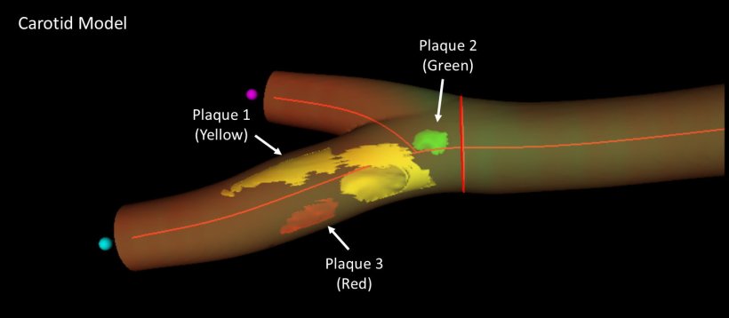
The burden, or quantity, of atherosclerosis in the carotid and femoral arteries is a well-established marker of cardiovascular risk and is highlighted as a key parameter in international clinical practice guidelines and expert consensus documents. There is therefore a recognized need for better and easy-to-use methods for measuring plaque burden that can be used as population screening tools.
The new imaging method was first validated and implemented in a study of almost 200 healthy participants with an intermediate cardiovascular risk from the Athero Brain: Head-to-Heart study, led by Dr. Valentín Fuster, Director General of the Centro Nacional de Investigaciones Cardiovasculares (CNIC). The method has now been incorporated into the PESA-CNIC-SANTANDER study, also led by Dr. Fuster, where it is being used to assess more than 4000 healthy individuals over a 9-year follow-up. The study, which started in 2010 and was recently extended until 2030, is one of the most important cardiovascular prevention studies in the world.
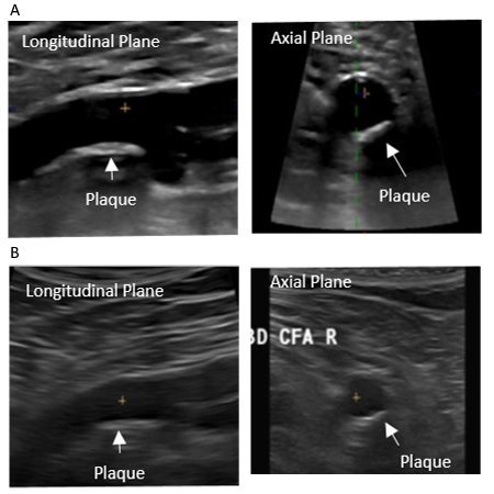
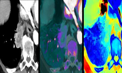
Image source: CNIC
The CNIC researchers partnered with Philips Ultrasound and Philips research Paris-Medisys to develop a new probe and software for real 3D ultrasound to facilitate exploration of the carotid and femoral arteries and speed up quantification of atherosclerotic plaque volume. As Dr. Fuster explained, “it is clear that traditional clinical evaluations based on measurements of cholesterol, blood pressure, blood glucose, and lifestyle habits cannot, on their own, accurately determine accumulated damage in the cardiovascular system, and without this crucial information we cannot take appropriate decisions to prevent acute events such as myocardial infarction or stroke.” The key to personalized prevention and treatment strategies, added Dr. Fuster, “is the ability to detect and quantify an individual’s accumulated cardiovascular damage, or atherosclerotic burden, using noninvasive imaging techniques.”
The newly validated 3D vascular probe incorporates 3D matrix technology, which underpins the most advanced 3D ultrasound techniques. CNIC Clinical Research Director Dr. Borja Ibáñez explained that the new technology allows simultaneous analysis by 2D and 3D ultrasound, includes all functionalities (color doppler, power-doppler, and contrast ultrasound), and is easily incorporated into daily clinical practice by technical and medical teams already experienced in ultrasound, emphasizing that “the integrated analysis software incorporates real 3D data processing.”
We now have a tool that can be used on-the-fly in an initial consultation, speeding up decision making – an important consideration in cardiovascular prevention, where time is of the essence
Beatriz López-Melgar
In addition to demonstrating the accuracy of 3D matrix ultrasound, the study demonstrates that the new technique takes just half the time needed by previous methods to obtain all the information required for the definition of carotid and femoral plaque burden, essential information for correct patient management.
For this reason, added Dr. Beatriz López-Melgar, “we believe that the development of ultrasound methods will contribute to the expansion of personalized medicine and the use of diagnostic imaging techniques, and will also help to ensure that improvements in patient care produce benefits for a larger sector of the population.” She concluded that, with the development of this technology, “we now have a tool that can be used on-the-fly in an initial consultation, speeding up decision making – an important consideration in cardiovascular prevention, where time is of the essence.”
Source: Centro Nacional de Investigaciones Cardiovasculares
17.03.2022