Image credit: Radiological Society of North America (RSNA)
News • 3D/4D visualization method
MRI volume rendering reveals pediatric heart defects in motion
With a new MRI technique that shows both heart tissue and blood flow simultaneously, physicians can see where heart defects occur and precisely plan to repair them, according to new research.
Their insights were published in Radiology: Cardiothoracic Imaging, a journal of the Radiological Society of North America (RSNA).
Researchers at Children’s Hospital of Philadelphia (CHOP) in Pennsylvania have developed 3D volume rendering methods for cardiac MRI that display complex structures within the heart and show how blood moves through them, much like ultrasound images but without the typical challenges of positioning angles. In their study, the researchers demonstrated how their methods guided treatment decisions in four young children who had complex congenital heart conditions.
In patients with holes in the heart structure or leaflets that don’t form a complete seal, we can now see the valve leaflets moving and identify exactly where a valve is leaking, which has not been possible with MRI before this technique
Matthew Jolley
Volume rendering is a computer graphics technique that creates 3D images directly from MRI scan data. It works by assigning colors and transparency to different tissue types based on how they appear in the MRI.
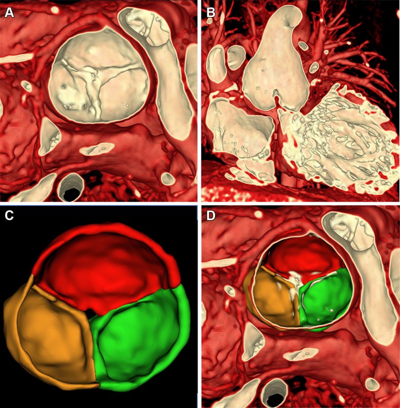
“Think of it like adjusting the settings on a photograph to highlight certain features,” said study coauthor Matthew Jolley, M.D., a pediatric cardiac anesthesiologist and cardiologist at CHOP and an associate professor at the University of Pennsylvania. “We developed specific settings that make heart muscle and heart valves visible while making blood and surrounding tissues transparent.”
The technique is particularly useful for observing blood flow through complex structures like valve leaflets—the flaps within the heart valves that are designed to open to allow blood to flow through, then close to form a tight seal and keep the blood from leaking backward in the wrong direction.
“In patients with holes in the heart structure or leaflets that don’t form a complete seal, we can now see the valve leaflets moving and identify exactly where a valve is leaking, which has not been possible with MRI before this technique,” Dr. Jolley said.
One patient in the study, a 4-year-old boy with a leaking and narrowed aortic valve, was being evaluated for valve repair or replacement surgery. The research team’s visualization tools showed the valve leaflets and a central jet of leakage, guiding the best surgical approach.
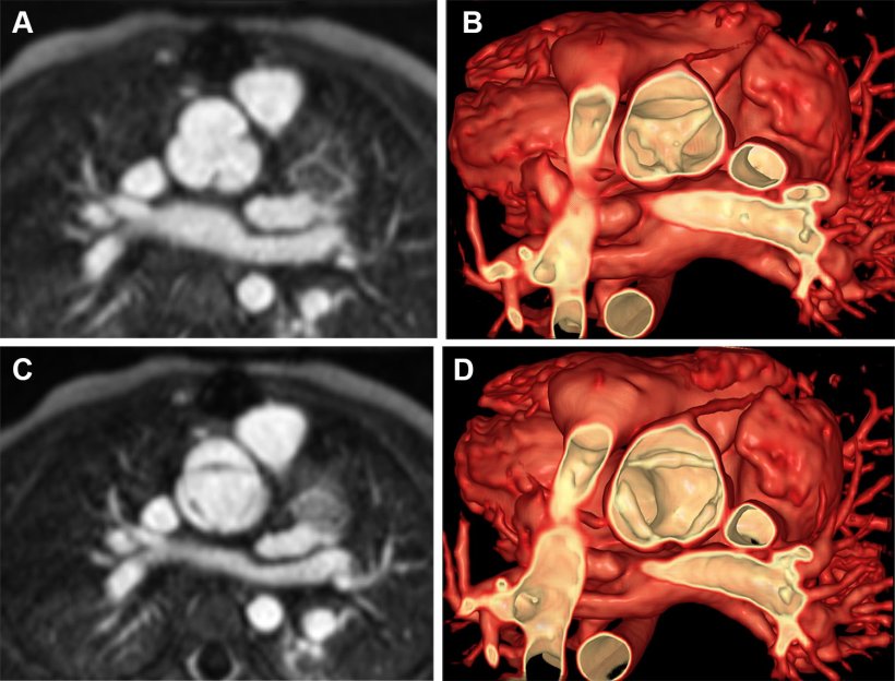
Image credit: Radiological Society of North America (RSNA)
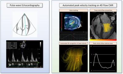
The team developed new ways to display blood flow on MRI, including lines showing the direction of flow and color-coded displays similar to those on Doppler ultrasound. While 3D ultrasound can also show tissue and flow together, it displays a smaller field of view, and the accuracy of flow measurements depends on the angle between the ultrasound beam and the blood flow direction, Dr. Jolley explained. CT can provide excellent anatomic images but cannot show blood flow, and it uses ionizing radiation. MRI provides high-quality flow images regardless of angles, and it does it without radiation—which is especially important for children who may need repeated imaging throughout their lives.
“Importantly, volume rendering is fast—generating visualizations nearly instantaneously—which is essential for 4D moving images where there is simply too much information to process using traditional manual tracing methods,” Dr. Jolley said.
Dr. Jolley said that the team sees these MRI visualization techniques as a complement to ultrasound rather than a replacement. “Our approach has limitations,” he said. “The quality of these visualizations depends heavily on the quality of the underlying MRI scan. Approaches like manual tracing can correct for image imperfections and is still necessary for certain analyses like computer simulations of heart function.”
The team was excited to find that their MRI-based images looked quite similar to 3D echocardiography with color Doppler, which doctors are already familiar with and rely on for evaluating heart valves.
From this work comes a suite of free cardiac image processing tools, SlicerHeart, developed using an open-source program called 3D Slicer. The research team has made the tools available for research and treatment in cardiovascular medicine, especially congenital heart disease, at SlicerHeart.org.
Source: Radiological Society of North America
13.02.2026