CT Perfusion of the Brain
The basics of the method and interpreting images
CT assisted dynamic perfusion imaging (perfusion CT, PCT) has evolved in recent years with the introduction of the multi-slice spiral technique, the use of study protocols with lower injection rates and improved evaluation programmes.
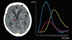
Fig. 1: Typical time/density curves after injection of a contrast mediumbolus in Perfusion CT. The density sequences are imaged (idealised view) in an arterial vessel (middle branch of the cerebral artery: blue), a venous vessel (confluence of sinuses: yellow) and in the cerebral parenchyma (thalamus: red). Note the typical staggered time between the arterial and venous time/density curves and the flattened
and slightly delayed density sequence in the parenchyma compared with the arterial curve
and slightly delayed density sequence in the parenchyma compared with the arterial curve
This article was first published in the VISIONS, issue 9/2006, a publication of Toshiba Medical Systems
03.07.2007