Sponsored • Innovation
The future of elastography rides on the shear wave
A practicing radiologist specialising in ultrasound, Pavlos Zoumpoulis MD PhD is also President and CEO of Diagnostic Echotomography, a day clinic based in Kifissia, Greece. The past President of the Hellenic Society of Ultrasound in Medicine and Biology spoke with our European Hospital team about his experiences with the next-generation in shear wave elastography on Mindray’s Resona 7 platform.
You are a leading advocate for elastography in ultrasound, calling it a revolution. What does that mean for you?
Pavlos Zoumpoulis: ‘As a radiologist, I’m familiar with all the imaging modalities but, early on, I focused on ultrasound, believing it had an exciting future. It seems like every five years there are new, revolutionary applications and innovations. The newest chapter in this long history of innovation is shear wave elastography, though elastography already has a long history of its own. The first technique was strain elastography using compression, which was useful for superficial organs such as the thyroid and the breast. Shear wave elastography brought a true innovation: a new technology that enabled us to apply this examination for tissue stiffness to more deeply situated organs, such as the liver, kidney, prostate, uterus, ovaries or pancreas. It also allows us to go beyond assessing tumours to evaluating diffused disease conditions. The most important application here is diagnosing liver fibrosis, the result of chronic liver disease.Shear wave elastography becomes an essential tool to study these conditions and, significantly, it can replace the need for other invasive examinations, such as needle biopsies.’
In your practice, compression or strain elastography is now part of history?
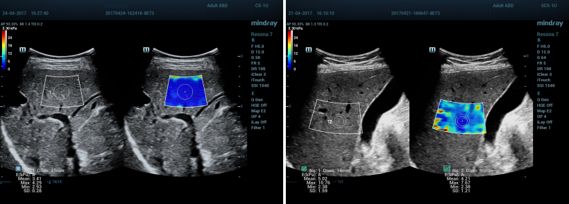
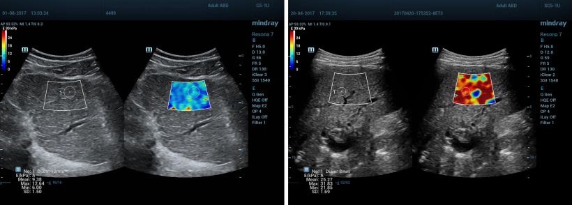
‘Using the strain technique of elastography we could not reliably get quantifiable results. Shear wave technology brings us this capability. Now we can calculate tissue stiffness by measuring it in units of kilopascal (kPa). We’ve always known that cancerous tissue will be stiffer than the normal tissue in an organ.Now, by scanning with Shear wave elastography, we can discriminate normal tissue from cancerous tissue and reliably report the result in measures of kilopascal. The quantification in kilopascal helps us to determine accurately the level of tissue stiffness associated with diffused liver fibrosis. I insist on the importance of reliability because, in the past, there have been problems with inter- and intra-observer variability of measurements using elastography. And this has been true for all the manufacturers, whether with a shear wave or a strain elastography capability. In our clinic we have been working with the Resona 7 for several months and it has proven to be highly reliable.’
Can the elastography chapter be closed, or do you still see an exciting future here?
There’s work to do in standardising measures
Pavlos Zoumpoulis
‘We are only at the beginning. With elastography we have already learned so much about disease in relation to tissue stiffness and we are going to discover even more, about the tissue characteristics, and content in fibrotic tissue or fat. Returning again to the liver, shear wave elastography has been widely adopted to assess diffused diseases, though there’s work to do in standardising measures. Different scales of measurements have been developed to determine the level of stiffness according to the cause of the disease, whether Hepatitis B, Hepatitis C or Cirrhosis due to alcoholism. We have seen rapid advances for ultrasound in musculoskeletal exams.Muscles and tendons are superficial, so they are easily visualised with ultrasound. Therefore, we can now apply shear wave elastography because, unlike strain elastography, it does not rely on compressing tissues, which results in more reliable measures of the stiffness. This becomes very important in evaluating the disease condition, or even to assess whether any tumours we identify are malignant or benign.’
What about paediatric diseases?
‘Unfortunately, there are chronic liver diseases that affect children. Yet, because children are smaller, fortunately it becomes easier to apply the benefits of shear wave elastography in examinations. Today we can reliably and non-invasively measure stiffness using ultrasound, rather than sending them for a liver biopsy procedure. We can also rely on these measures to grade the condition of the disease. The examination becomes even easier with the new technology, for the doctor, and especially for the patient who avoids both pain and complications because it’s only an ultrasound exam.’
You recently opened a training base at EchoMed Centre, in cooperation with Mindray. What has been your experience with this?
‘Not every elastography application manufacturer has a commitment to training physicians. Mindray is one that does believe education goes hand-in-hand with its technology and that physicians need to understand how to apply the technologies as an essential step. For those with the skills and knowledge, the Resona 7 makes shear wave elastography easier to apply, but this is not to say that shear wave elastography is so simple anyone can do it. The program we offer is intensive, 18 hours of training covering theory and practice across all organs. It is also highly interactive. The participants come from all over the world – not only Europe, but Asia and North America as well. So far, I can tell you that much; it’s international, intensive and very interactive.'
Profile:
Pavlos Zoumpoulis MD PhD is practicing radiologist specialising in ultrasound as well as President and CEO of Diagnostic Echotomography, a day clinic based in Kifissia, Greece. As President of the Hellenic Society for Ultrasound in Medicine and Biology (HSUMB) he organized for the first time ever 2015 in Greece the EUROSON, the European Congress for Ultrasound.
14.11.2017