Ars Electronica Center
Virtual journey through the heart
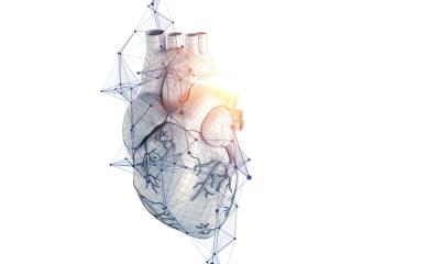
Medical research and art sometimes meet at their finest: experts from the Fraunhofer Institute for Medical Image Computing MEVIS in Bremen produced a three-dimensional movie, showing the human heart in full action. The organ beats and pumps, and special techniques visualize the dynamic flow of blood in the vessels. The sequence is part of a new interactive three-dimensional experience to be presented by the “Ars Electronica Center” in the “Deep Space 8K” experience in Linz today.
The Ars Electronica Center and its annual festival belong to the leading showrooms and exhibition centers for digital culture. As a museum of the future, it shows the visitors how nascent technologies, still in development, could shape the daily life of the future – from the workplace to leisure time and art.
The Deep Space projection hall is a part of the Center and can display three-dimensional images, movies, and animations in extremely high resolution. Deep Space has been refitted with new 8K technology that improves image sharpness and color intensity. Moderators accompany the experience with live expert commentary.
For the reopening, the Ars Electronica Center has developed a new three-dimensional program for the Deep Space adapted to the optimized projection technology. “The Universe Within” offers an interactive journey through a three-dimensional visualization of our body and shows how organs, bones, muscles, and blood vessels appear and function. Fraunhofer MEVIS contributed a central element to the exhibit – a three-dimensional sequence of the human heart.
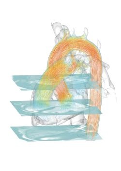
Images from a CT scanner build up a three-dimensional block. Gradually, the ribs, diaphragm, and beating human heart can be recognized. The camera perspective changes, and a single image layer comes to the foreground, showing how heart valves open and close with the heartbeat. Refined algorithms visualize the blood flow based on an MR image that appears thereafter.
Hundreds of colored sparks fly in rhythm with the heartbeat through the vessels and heart chambers and visualize the areas where blood flow is particularly fast and forceful. When not accompanied by live commentary, a soundscape composed specifically for the sequence can be heard.
The three-minute sequence is based exclusively on real medical data gathered by the Institute’s MRI scanner. The CT data were provided by the University Clinic in Marburg. Fraunhofer scientists created the movie using “MeVisLab”, a software package for developing medical image processing assistance systems for physicians. Experts and industrial partners around the world currently use MeVisLab.
The researchers used sophisticated volume rendering methods to create the aesthetical images. The blood flow sequence is a product of a new method, in the process of being applied to the clinical routine in Bremen, called particle-based flow visualization. The concept stems from computer simulations for wind channel experiments. Fed with the MRI data and adjusted to the human body, it can now visualize blood streams in vascular system.
Other techniques used for the video have already proven themselves in the clinical routine. Doctors can use software assistants to view specific images of the data set that present important details, such as heart valves, particularly well.
The heart sequence is not the first joint project between Bremen and Linz. At the Ars Electronica Festival in 2013, Fraunhofer MEVIS showcased “Poking Florian,” an interactive installation that visualizes the nerve fibers and their function in the human brain. Other cultural institutions also profit from exhibits designed at Fraunhofer MEVIS. Recently, “Image Man” was installed in the Universum Science Center in Bremen. The exhibit is a life-size figure containing ten segments that present the possibilities of different imaging methods, such as X-ray, CT, and MRI.
The experts work not only on medical videos and hands-on exhibits based on real medical data; they also develop applications for virtual reality glasses. In the future, their programs could help train medical personnel and explain information to patients.
Watch 2-D movie on YouTube: https://youtu.be/0B0d0fPVcKI
Source: Fraunhofer MEVIS - Institut für Bildgestützte Medizin
07.08.2015