Image source: BCS/University of Sheffield/VirtuHeart
News • 3D vascular models
'Virtual blood vessel' technology could improve heart disease care
Patients with heart disease could benefit from less extensive interventions thanks to cutting-edge technology that creates 3D computer models of blood flow through the heart's arteries, according to research presented at the British Cardiovascular Society in Manchester.
When the research team trialled the "VirtuHeart" technology with doctors treating heart attack patients, they found that using it would have changed the treatment of more than 20% of patients. In many cases, it would have led to fewer patients undergoing an invasive procedure such as having a stent fitted.
By giving doctors a clearer picture of a patient’s arteries, the research funded by the British Heart Foundation (BHF) showed that VirtuHeart could help more heart patients to get the right treatment for them, free up doctors’ time and better meet demand on heart care services.
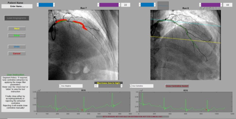
Image source: BCS/University of Sheffield/VirtuHeart
The researchers are currently investigating the impact this technology could have if it was used widely in the NHS, including the effect it might have on waiting lists. They hope that it could be in use in as little as three years. Dr Hazel Arfah Haley, Interventional Cardiologist at Sheffield Teaching Hospitals NHS Foundation Trust, led the study. She said: “By giving doctors a better understanding of what is happening inside their patient’s blood vessels, we’ve shown that this technology has the potential to help improve how we assess and treat heart disease, ensuring patients have the treatment that best meets their needs. Our team are also investigating whether VirtuHeart could improve treatment for people with another common heart condition called angina, helping to make sure that even more patients get the treatment they need first time around.”
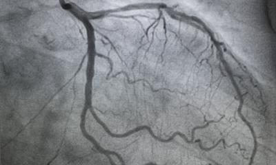
According to data from the Bristish Cardiovascular Intervention Society (BCIS), there are up to 250,000 coronary angiograms performed in the UK every year - a test which allows doctors to look inside a patient’s coronary arteries (which supply the heart muscle with blood) and check for blockages. This is one of the first tests that patients admitted to hospital with a heart attack will undergo and helps doctors plan treatment to restore blood supply to the heart muscle. But angiograms can be hard to interpret when an artery is only partly blocked, and this can make it challenging for treatment decisions to be made, particularly when doctors are managing patients with complex heart disease.
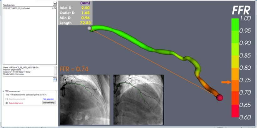
The innovative technology, developed by researchers at the University of Sheffield, re-creates another invasive but underused test called a Fractional Flow Reserve (FFR) in which doctors insert a special wire into arteries to calculate how well blood is flowing. FFR is underused for several reasons including time pressure, availability, complex anatomy, and operator’s familiarity.
Recommended article
Article • Predicting plaques
Exposing the secrets of the heart
Coronary interventions often rely more on art than science as the decision to treat a patient tends to be based on what clinicians can see, a subjective interpretation of cardiac imaging. Two new techniques have emerged for cardiovascular diagnostics that are enabling software to help surgeons and cardiologists measure, and thereby better manage cardiac disease.
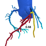
Using only the images from a patient’s angiogram, VirtuHeart works as a “virtual FFR” and creates computer models of their blood vessels, allowing doctors to calculate blood flow and find out more about the extent of blockages.
Professor Sir Nilesh Samani, Medical Director at the British Heart Foundation, said: “There are around 100,000 hospital admissions each year due to heart attacks, mostly caused by the coronary arteries becoming narrowed and blocked by a build-up of fatty plaques. Technologies like VirtuHeart show real promise to improve treatment as well as reducing unnecessary interventions, expense and complications and freeing up clinicians’ time to treat other patients.”
The study involved 208 patients who were admitted to hospital with an NSTEMI – a type of heart attack where the affected coronary artery isn’t completely blocked. All of the patients had their coronary arteries reconstructed using VirtuHeart. After the patients had been treated, the researchers revealed the virtual blood vessel models to their doctors. They found that using the technology would have changed how doctors treated 46 patients (22%). Of these, 21 patients who had an invasive procedure such as a stent would have instead been treated with medication only if the technology had been used to plan their treatment. Overall, using VirtuHeart to plan treatment would have led to 42 fewer stents being fitted – a decrease of 18%.
The VirtuHeart system was developed by the Mathematical Modelling in Medicine research group in the department of Infection, Immunity and Cardiovascular Disease at the University of Sheffield, in partnership with the Insigneo Institute and Sheffield Teaching Hospitals NHS Foundation Trust.
Source: British Cardiovascular Society
09.06.2023