Ultrasound in appendicitis re-evaluation
Boosting diagnostic accuracy and the decrease of negative appendectomy rate
Dr Hyuk Jung Kim
Researchers have found that ultrasound has an important role to play in re-evaluating patients with findings of acute appendicitis. Although CT is now accepted as an excellent system to diagnose acute appendicitis, misdiagnosis still occurs in routine practice, prompting a team from South Korea to conduct a study to prospectively evaluate the additional diagnostic value of ultrasound (US) re-evaluation for patients with equivocal CT findings of acute appendicitis.
Further diagnostic value
The research team, led by Dr Hyuk Jung Kim from the Department of Radiology at Bundang Jesaeng General Hospital, found that if there are equivocal CT findings of acute appendicitis, US re-evaluation can be used to improve diagnostic accuracy and further decrease the negative appendectomy rate.
The team focused on 869 consecutive patients with suspected appendicitis who were referred for CT. The likelihood of appendicitis was prospectively placed in five categories with US re-evaluation recommended for patients in the ‘equivocal appendix’ and those who probably did not have appendicitis based on CT findings. The final decision to proceed with US re-evaluation for equivocal cases was made by the treating physician on the basis of clinical and laboratory findings.
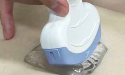
‘After adding US re-evaluation,’ Dr Kim said, ‘the overall negative appendectomy rate in our institution decreased from 3.4% to 2.3%.’ The team also found a number of additional diagnostic values from ultrasound re-evaluation for patients with equivocal CT findings of acute appendicitis. ‘Graded compression ultrasound was helpful for distinguishing acute appendicitis from dilatation due to fluid or faeces,’ he said, adding: ‘ultrasound probe-induced tenderness over the appendix was also an important finding to differentiate between non-inflamed dilated appendix and inflamed appendix. ‘In addition, the spatial resolution of high-frequency ultrasound image provides more detailed visualisation of appendiceal wall than that of CT.’ A further benefit for such patients, he said, is that US re-evaluation can be used to improve diagnostic accuracy and further decrease the negative appendectomy rate without increasing the perforation rate.
In his department they usually recommend APCT as a primary modality to evaluate the appendix for adults and young patients, except pregnant women and relatively thin child patients where ultrasound is recommended as the primary modality. US use in this context has benefits for both clinicians and patients: ‘When patients have equivocal CT findings of appendicitis, the clinician should make a decision whether to undergo surgery based on experience and the patient’s clinical manifestation. If the patient does not undergo surgery, Dr Kim pointed out that s/he will be kept under close hospital observation. ‘In this case, the risk of delayed complication such as perforated appendicitis will exist.
‘In our study, after implementing US re-evaluation, the appendiceal perforation rate is decreased as well as the negative appendectomy rate. For patients, it could decrease the length of hospital stay and cost of hospitalisation,’ he added. A number of other factors need consideration when using US in this way, in particular ensuring that operators
are properly trained because ultrasound diagnosis is operatordependent, Dr Kim explained. ‘Patient-related factors such as age, body habitus and appendix location are important influencing factors. ‘Evaluation of the appendix tip is an especially challenging part of a US exam, because in most patients the appendix tip is located in the deep pelvis. Paying close attention to the evaluation of the appendix tip can improve diagnostic accuracy.’
Dr Hyuk Jung Kim is section chief of abdominal imaging in the Department of Radiology at Bundang Jesaeng General Hospital, South Korea. He has 12 years of experience in abdominal imaging. Ji Ye Sim, a fourth-year resident at the hospital’s Department of Radiology, played a key role in the research.
Report: Mark Nicholls
12.11.2013
More on the subject: