Ultrasound 60 years later
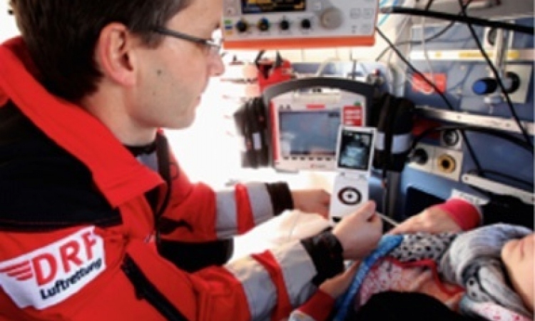
Through the miracle of modern-day ultrasound, we are able to see – in three dimensions and in real time – the functioning of arteries, veins and the many sophisticated structures of the heart. While most think of ultrasound technology as it relates to grinning parents getting a first glance of their baby in the womb, cardiovascular care is being revolutionised by advances in ultrasound technology.

This year, we celebrate 60 years of ultrasound in the medical field. Its use in the medical community began in 1953, when physician Inge Edler and engineer C Hellmuth Hertz produced the first echocardiogram of the heart in Sweden. Incredibly, they did this by using an industrial-sized ultrasonic tool from a neighbouring shipyard that was being used to detect cracks in the metal of ships.
In 1956, Japanese physicians used the Doppler Effect (the change in wave frequency relative to distance) to examine cardiac motion. Following the Japanese discovery, Doctors John Reid and John Wild in Minnesota developed the first ultrasonic scanner for clinical use.
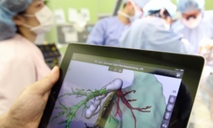
Today’s premium ultrasound systems meld live 3-D images with tools like 2-D Intracardiac Echo (ICE) capability that present very clear, rich images to aid clinicians in a host of intricate cardiovascular repairs that are increasingly done in a minimally invasive way. Procedures to treat conditions such as valve insufficiency or defects in the septal walls are made possible largely due to the ability of today’s ultrasound technology to provide the road map.
The next 60 years of ultrasound and other evolving imaging technologies are sure to bring us many more innovations aimed at improving healthcare in a variety of clinical settings around the world.
05.11.2013