Image source: UCL
News • Hypertrophic cardiomyopathy imaging
Combining cDTI and CMR to detect deadly heart condition
Combining two types of heart scan techniques could help doctors to detect the deadly heart condition hypertrophic cardiomyopathy (HCM) before symptoms and signs on conventional tests appear, according to a new study led by researchers at University College London (UCL).
The research, funded by the British Heart Foundation and published in the journal Circulation, opens the prospect of treating the condition at the earliest stages. Being able to detect HCM earlier than ever before will also assist trials investigating gene therapies and drug treatments aimed at stopping the disease developing in those at risk.
HCM is an inherited condition that affects around 1 in 500 people in the UK. It causes the muscular walls of the heart to become thicker than normal, affecting how well the heart can pump blood around the body. It is a leading cause of heart failure and sudden cardiac death.
The ability to detect early signs of HCM could be crucial in trials testing treatments aimed at preventing early disease from progressing or correcting genetic mutations
George Joy
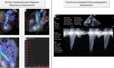
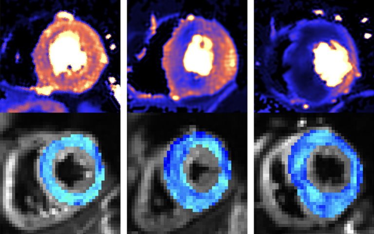
Researchers from UCL, Barts Heart Centre and University of Leeds studied the hearts of three groups: healthy people, people who already had HCM, and people with an HCM-causing genetic mutation but no overt signs of disease (no heart muscle thickening). To do this, they used two cutting-edge heart scanning techniques: cardiac diffusion tensor imaging (cDTI), a type of MRI scan that shows how individual heart muscle cells are organised and packed together (the heart’s microstructure), and cardiac MRI perfusion (perfusion CMR), which detects problems with the small blood vessels supplying the heart muscle (microvascular disease).
The scans showed that people with overt signs of HCM have very abnormal organisation of their heart muscle cells, and a high rate and severity of microvascular disease compared to healthy volunteers. Crucially, the scans were also able to identify abnormal microstructure (muscle cell disorganisation) and microvascular disease in the people who had a problematic gene but no symptoms or muscle thickening. They found that 28% had defects in their blood supply, compared to healthy volunteers. This meant that doctors were able to more accurately spot the early signs of HCM developing in patient’s hearts.
Recommended article

Article • High tissue contrast, spatial detail, complete tissue characterisation
MRI shows cardiac diagnostic value
Cardiovascular magnetic resonance (CMR) imaging has become faster, simpler and more widely available in recent years because it has evolved to deliver effective assessment and diagnosis of a range of heart conditions with expanding guideline indications.
The first drug to slow HCM progression – mavacamten – has recently been approved for use in Europe and will allow doctors to reduce the severity of the disease once symptoms and muscle thickening have appeared. Genetic therapies are also in development which could prevent symptoms entirely by intercepting HCM development at an early stage.
Perfusion CMR is already being used in some clinics to help differentiate people with HCM from other causes of muscle thickening. The researchers think that these revolutionary new therapies, combined with cDTI and perfusion CMR scans, give doctors the best ever chance of treating people at risk of HCM early enough that the condition never develops.
Dr George Joy, who led the research with Professor James Moon and Dr Luis Lopes (all UCL Institute of Cardiovascular Science), said: “The ability to detect early signs of HCM could be crucial in trials testing treatments aimed at preventing early disease from progressing or correcting genetic mutations. The scans could also enable treatment to start earlier than we previously thought possible. We now want to see if we can use the scans to identify which patients without symptoms or heart muscle thickening are most at risk of developing severe HCM and its life-changing complications. The information provided from scans could therefore help doctors make better decisions on how best to care for each patient.“
Dr Luis Lopes (UCL Institute of Cardiovascular Science), senior author of the study, said: “By linking advanced imaging to our cohort of HCM patients (and relatives) with extensive genetic testing, this study detected microstructural abnormalities in vivo in mutation carriers for the first time and was the first to compare these parameters in HCM patients with and without a causal mutation. The findings allow us to understand more about the early subclinical manifestations of this serious condition but also provide additional clinical tools for screening, monitoring and hopefully in the near future for therapeutic decision-making.”
Source: University College London
20.07.2023