Progress
Scientific meetings held since 1998 at Alpbach, Germany, have attracted the sponsorship of leading associations and companies such as the Philip Morris External Research Programme, the Donors Association of German Science, Swiss National Fund, the German Heart Centre Foundation, Berlin, and Philips Medical Systems. At the 4th Alpbach Meeting, which focused on Magnetic Resonance, Contrast Mechanisms and Molecular Imaging of Coronary Artery Plaques, Daniela Zimmermann spoke with Professor Eckart Fleck of Berlin's German Heart Centre (Deutsches Herzzentrum Berlin) about the constantly increasing importance of this event and its current aims
Cardiologists, radiologists, physicists, vascular biologists, and other specialists arriving at Alpbach, Germany, could not fail to admire their surroundings. ‘Alpbach is very beautiful and has a long tradition as a place for universities and the European Forum, which seemed a particularly attractive combination for our meetings. It provides the atmosphere you need to concentrate on topics, combined with a chance to relax in pleasant surroundings,’ said Professor Fleck, explaining the choice of venue.
Here, the present and future issues to be faced in cardiology are discussed, centred on two approaches: ‘First, the enhancement of diagnostics to detect potential dangers at an early stage - meaning not just in time,’ the professor explained. ‘Second: What treatment? If one bears in mind that arteriosclerosis will not only continue to persist but also increasingly prove a general health problem, then of course diagnostic considerations of this nature are of paramount importance. In 2020 cardiac diseases, and particularly coronary heart disease, will be the number one cause of death, worldwide, and not just in industrialised countries as at present. At the moment, infectious diseases hold that position, but, as pointed out, this will change worldwide, mainly because of changing dietary habits and that people are living increasingly longer. Arteriosclerosis will ultimately affect us all, if we live to be old enough.’
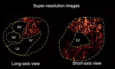
At the Alpbach meeting the convergence of imaging and treatment was constantly demonstrated. ‘The critical areas of interest to us are plaque formations, that is deposits on local vascular wall sites, something that we cannot really detect from the outside. We need in vivo information about this. To date, imaging is conducted within the vessel itself; not only angiographically showing the internal volume but attempts are made to depict the wall. This is possible, for example with ultrasound and new modalities that can produce slice images of adequate resolution. As such, this could diagnose millimetre structures in the vascular walls.
This is possible using CT, but there are major problems - even to be able to see wall thickness and calcification. Conversely, by using MRI one can determine the different quality of tissue and obtain a resolution of practically microscopic quality, without using radiation. Imagine how exciting it is to conduct histology in a body that is completely penetrated by light. This is possible not only thanks to the resolution but also by marking those sites that you seek. The combination of these two approaches is the focus of the Alpbach meeting. The scientists ponder what are the potential binding possibilities and what individual proteins or other enzymes are known and could be considered as a target for specific binding sites, because this has implications for the combination of contrast media. In future it is even conceivable - should it be possible to identify such proteins and enzymes - to also use these for treatment, because you can see where and how the therapeutic agent acts at a specific site. This conjures up futuristic images and is based on the assumption that we work at obtaining high resolution of local visualisation and on identification of receptor proteins.’
Multidisciplinary research such as this, drawing together biologists and chemists as well as medical practitioners, must be exciting. ‘Yes, it is,’ Prof. Fleck confirmed. ‘The purpose of our workshop is to try to have completely different approaches and viewpoints bounce off each other, for scientists to engage in cross-talk, so that one group grasps what others need and vice versa. Because one cannot assume that a chemist, who is thoroughly conversant with special binding issues, will understand what he should really do if he is not made aware of the context and cannot understand what it’s all about - and, of course, vice versa. What do you do with an image when you do not know how it was generated and thus misinterpret? So we must know relatively a lot about the local circumstances, not only about diagnosis of disease. We should know what specific particles are used and whether these really do bind as selectively as desired. Or is the receptor - a good early marker of inflammation and is expressed in large quantities in an area - really capable of binding, and, assuming the answer is yes, just what that means. If you have a molecule with an adequate number of receptors that you can bind to the vessel wall, you should be able to see this site well - if enough contrast medium is given. Then there are special contrast media that give good signals in a magnet - paramagnetic beads. These are small, non-magnetic iron particles. If we could bind these beads to specific molecules, which, in turn, would dock on to receptors, then you would have created a permanent link at this site, which could be visualised with MRI - assuming that a sufficiently large quantity is available. If all that is possible, you could localize a particular site.’
Professor Fleck agreed that the MRI-PET scanner would be a useful machine is such a situation, ‘... because it appears to be much easier to establish the special binding forms I spoke of earlier - in nuclear imaging. It would work very well with PET, but the only drawback with PET is that it does not at all have the kind of local resolution we need. If MRI could be used here, for example, in combination with specific contrast media, then one would probably not need PET anymore. Perhaps PET can be used as a general investigation system, while selectively using MRI to generate the high resolution needed. However, we believe that in future using MRI alone we will be able to visualise specific binding forms and thus achieve the requisite precision.
'We have been aware for years that conventional diagnostic modalities were not enough and that we would have to achieve higher quality using less invasive approaches. Our insights to date come from relatively highly invasive procedures. This is something that we would like to change - particularly in preventive medicine. We do not want to subject healthy, or potentially healthy, individuals to a battery of tests that, in some cases, could cause disease themselves. We therefore need data that will, a priori, assure equal, if not better, precision. Indeed, medicine has always explored such investigation procedures, be it nuclear medicine procedures or ultrasound, etc. But all the procedures mentioned suffer the drawback that local resolution is not good enough. For that reason we began working in this field as soon as the technology- fast computers for image reconstructions and strong magnets that can acquire the required signal - became available.’
03.08.2006