Image source: Adobe Stock/Rasi
News • HCC in vitro research
Organoids give new clues to liver cancer progression
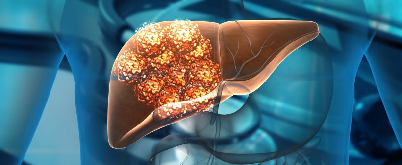
A team of researchers from the College of Design and Engineering, the N.1 Institute for Health and the Cancer Science Institute of Singapore at the National University of Singapore has recently engineered in vitro tumour models to better understand the crosstalk between liver cancer cells and their microenvironment.
Using lab-grown mini liver tumours co-cultured with endothelial cells – these are cells that form the lining of blood vessels – to conduct their study, the research team investigated the role of endothelial cells in liver cancer progression. The team’s results were published in the journal Biomaterials.
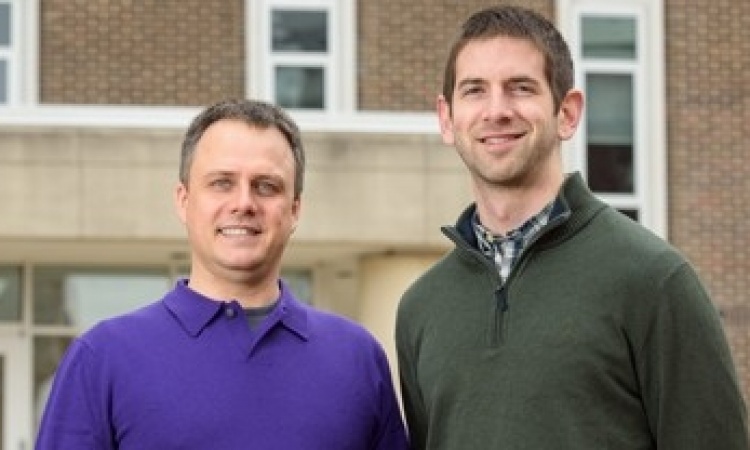
“The conventional understanding is that endothelial cells are structural cells that form blood vessels. Our latest findings suggest that these cells also give ‘instructions’ to liver cancer cells to increase the production of a protein called CXCL1, which is associated with poor survival outcome in liver cancer patients,” explained Assistant Professor Eliza Fong, who led the research study.
CXCL1 is a type of chemokine, which are signalling proteins secreted by cells to regulate the infiltration of different immune cells into tumours. Hence, these molecules affect tumour immunity and may influence therapeutic outcomes in patients. “Our results pave the way for new therapeutic targets to control tumour development, and further our team’s understanding of the mechanisms behind the progression of liver cancer,” Dr. Toh Tan Boon added, who is also a key member of the research team.
We found that the ‘communication’ between liver cancer cells and endothelial cells contribute to the generation of macrophages [...], potentially creating a very conducive microenvironment for tumour expansion
Edward Chow
Hepatocellular carcinoma (HCC) is the sixth most commonly occurring cancer and remains the second leading cause of cancer worldwide. While several therapeutics have been approved in the recent years to treat advanced HCC, the impact of anti-angiogenic treatment on the overall survival of HCC patients remains elusive. Previous studies have shown that tumour growth is facilitated by a biological process called angiogenesis (the development of blood vessels) which provides the tumour with oxygen and nutrients to grow. Unfortunately, the benefits of angiogenesis inhibitors are only temporary, following which the tumour resumes growth.
To find new therapeutic targets to better control tumour development, the NUS team decided to study the role of endothelial cells in cancer development using engineered in vitro tumour models. Unlike previously reported models that rely on the use of immortalised cancer cell lines, the NUS team incorporated patient-derived xenograft organoids and endothelial cells in their new model, which led the team to their significant discovery. “We found that the ‘communication’ between liver cancer cells and endothelial cells contribute to the generation of macrophages. These immune cells are pro-inflammatory and pro-angiogenesis, potentially creating a very conducive microenvironment for tumour expansion,” explained Associate Professor Edward Chow, also a key member on the research team.
This latest study, which is a continuation from their previous work in 2018, highlights the significance of setting up such complex co-cultures to better understand the liver cancer milieu. Importantly, these co-culture models may be useful for drug development studies looking to target liver cancer and can also serve as valuable platforms to better understand how inflammation is promoted in liver cancer, and how it contributes to cancer progression.
Moving forward, the research team hopes to leverage their practical knowledge to set up such co-cultures and extend this area of expertise to other cancer types. Their research work will provide an enhanced road map for the study of cancer-endothelial crosstalk.
Source: National University of Singapore
02.06.2022


