On its first test path
The prototype of the open X-ray system ORBIT generates detailed 3-D images

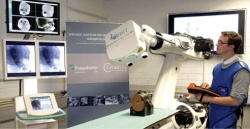
In the summer of 2011 European Hospital first presented ORBIT, a joint project by the Fraunhofer Institute for Production Systems and Design Technology (IPK), Charité Universitätsmedisin Berlin and Ziehm Imaging, and funded by the German Ministry of Education and Research (BMBF).
Today, the project director Professor Erwin Keeve PhD-Eng is proud to announce that the system has generated the first 3-D images: ‘We are very happy with these initial results, which motivate us to enter the next funding phase and move ahead towards clinical use.’
The second prototype is most likely to be installed in the state-of-the-art robotics operating theatre that he is currently setting up at Charité. ‘If everything runs according to schedule we will be able to demonstrate clinical usability of ORBIT by mid-2015,’ he says. If this pans out, the system, which is designed for minimum impact on surgical procedures and routine use in operating theatres, could be launched in 2016 for use in shock rooms, for example.
The clinical significance of a device like this was recently highlighted by a study based on the German trauma registry, which indicated that an immediate whole-body CT might significantly reduce mortality among polytrauma patients.
‘The advantages of ORBIT,’ Professor Keeve points out, ‘are the facts that, unlike a CT, it can be installed in the shock room itself and that it offers unrestricted access to the patient during image acquisition. Moreover, ORBIT is much simpler and faster to set-up for each individual exam.’
05.11.2013


