News • Renal function deterioration
MRI could predict kidney disease before it develops, study suggests
Mount Sinai researcher recognized for innovative MRI research that could improve prediction of kidney function decline after nephrectomy

Image credit: Mena Shenouda
An investigator at the Icahn School of Medicine at Mount Sinai has received international recognition for innovative imaging research that may help physicians identify patients at increased risk of chronic kidney disease (CKD) before they undergo surgery for kidney tumors.
Mira Liu, PhD, a postdoctoral fellow at the Mount Sinai BioMedical Engineering and Imaging Institute, received the prestigious W.S. Moore Award for Original Clinical Research at the 2026 International Society for Magnetic Resonance in Medicine & International Society for MR Radiographers & Technologists Annual Meeting & Exhibition. The award recognizes early-career investigators’ work for exceptional original research contribution in the clinical science of magnetic resonance (MR) with an emphasis on application of established MR methodologies with innovation, scientific rigor, and clinical relevance.
The award honors Dr. Liu’s article, “Multiparametric MRI Predicting Renal Function Deterioration & Chronic Kidney Disease Development in Patients Undergoing Nephrectomy for Renal Masses: A Pilot Study,” published in the Journal of Magnetic Resonance Imaging.
The study investigated whether advanced multiparametric magnetic resonance imaging (MRI) performed before kidney tumor surgery could help identify patients at higher risk for kidney function decline and chronic kidney disease after nephrectomy, the surgical removal of part or all of a kidney. “Patients undergoing surgery for kidney tumors often face uncertainty about how their kidneys will function afterward,” says Dr. Liu. “We wanted to investigate whether information already captured through advanced MRI could provide a more complete picture of kidney health before surgery and help support more personalized treatment decisions.”
That hidden stress may help explain why some patients are more vulnerable to kidney disease afterward, even when their kidney function initially appears normal
Mira Liu
Nephrectomy can be lifesaving, but some patients experience reduced kidney function afterward. Currently, physicians have limited tools to reliably predict which patients may be most vulnerable to long-term kidney complications.
In the pilot study, 43 patients underwent specialized research MRI scans before surgery in addition to their standard clinical imaging. The researchers used advanced MRI techniques to evaluate kidney blood flow, oxygen utilization, inflammation, filtration, and microscopic tissue characteristics. They then compared those findings with kidney function one year after surgery.
The researchers found that MRI measurements taken before surgery could help predict both kidney function decline and the future development of chronic kidney disease. Combining MRI-derived biomarkers with standard clinical assessments and blood-test results significantly improved the ability to identify patients at higher risk.
“What makes this work especially exciting is that it suggests MRI may reveal how hard the kidneys are already working to maintain healthy function before surgery takes place,” says Dr. Liu. “That hidden stress may help explain why some patients are more vulnerable to kidney disease afterward, even when their kidney function initially appears normal.”
The findings also highlight the broader potential of quantitative imaging to provide insights into organ function, not only anatomy, and to support more precise risk assessment before treatment. “This research demonstrates the growing power of quantitative imaging to support precision medicine,” says Octavia Bane, PhD, Assistant Professor of Diagnostic, Molecular and Interventional Radiology at the Icahn School of Medicine at Mount Sinai, a co-author and co-mentor of Dr. Liu. “By combining advanced MRI with clinical information, we may eventually be able to better personalize treatment plans, monitor high-risk patients more closely, and help protect long-term kidney health.”
The investigators emphasized that the study does not suggest patients should avoid surgery for kidney tumors. Instead, the goal is to help physicians identify patients who may benefit from additional kidney-protective strategies and closer follow-up care after treatment.
The study also revealed a surprising pattern: both poorly functioning kidneys and kidneys that appeared to be working exceptionally hard before surgery were associated with a greater risk of future kidney disease. Researchers believe that this may reflect reduced “kidney reserve,” meaning some kidneys may already be under strain even before surgery occurs.
“Our hope is that this work encourages further research into how MRI can provide a more complete understanding of organ health,” said Dr. Liu. “Ultimately, we want to help patients and care teams make more informed decisions using tools that are already part of clinical care.”
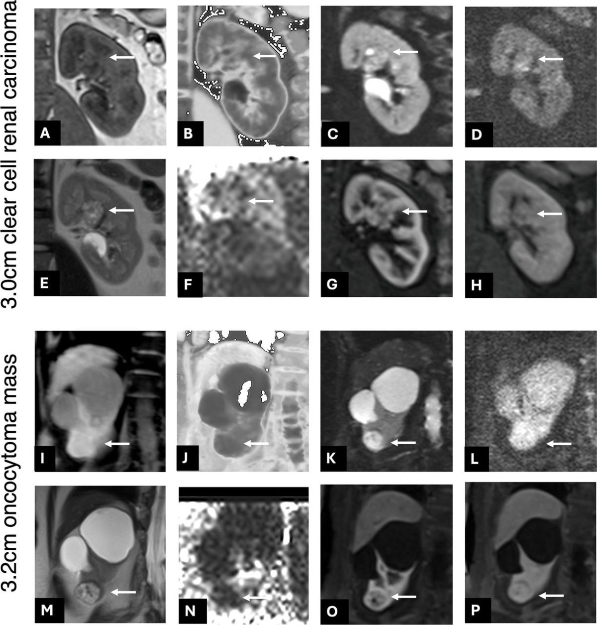
Image source: Liu MM, Bane O, Mu X et al., Journal for ImmunoTherapy of Cancer 2026 (CC BY-NC 4.0)
The research team plans to validate the findings in larger patient populations across multiple medical centers and hopes to develop a practical clinical-imaging risk score that could eventually be incorporated into routine presurgical evaluations.
In related recent research published in the Journal for ImmunoTherapy of Cancer, the investigators also found that similar presurgical MRI measurements correlated with features of tumor biology and the tumor immune environment. These findings raise the possibility that MRI could eventually provide insights into cancer behavior and kidney health simultaneously.
“We are delighted to see Dr. Liu recognized with the W.S. Moore Award for this innovative and impactful research,” says Sara C. Lewis, MD, Professor of Diagnostic, Molecular and Interventional Radiology at the Icahn School of Medicine at Mount Sinai who is co-mentor of Dr. Liu and senior author of the study. “Her work reflects the promise of advanced imaging to provide deeper insight into kidney health and patient risk, while advancing a more personalized approach to care for patients undergoing kidney tumor surgery.”
Source: Mount Sinai School of Medicine
14.06.2026