Image credit: Radiological Society of North America (RSNA)
News • Heart failure diagnostics
AI-enhanced MRI enables single-shot imaging of cardiac cycle
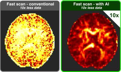
AI-enhanced single-shot cine MRI produces superior image quality and provides ventricular measurements comparable to conventional cine MRI.
This is according to new research published in Radiology: Cardiothoracic Imaging, a journal of the Radiological Society of North America (RSNA).
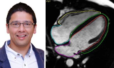
Accurate assessment of left ventricular function is essential for guiding treatment decisions, monitoring therapeutic responses and predicting outcomes in patients with heart failure. Cardiac MRI is the standard modality for evaluating left ventricular function and is traditionally performed using conventional cine sequences known as balanced steady-state free precession. Cine MRI captures moving images and effectively visualizes dynamic bodily processes in real time. However, it requires patients to hold their breath multiple times during the scan, which can be difficult for those with arrhythmia, or irregular heartbeat.
The AI-CS framework offers a promising alternative for cardiac MRI examinations in the clinical setting, where long acquisition time remains a major challenge
Nan Zhang
“For patients with severe arrhythmias, breath-holding can be particularly challenging, often resulting in compromised image quality, exam failure or inaccurate assessments,” said lead researcher Nan Zhang, M.S., supervisor radiologic technologist, Department of Radiology, Zhongshan Hospital of Fudan University in Shanghai. Single-shot cine sequences address this challenge by capturing the entire cardiac cycle in just two heartbeats, thereby shortening the breath-hold duration and significantly reducing the impact of arrhythmias on image quality.
In the study, Zhang and colleagues evaluated the feasibility of using deep-learning–enhanced Compressed SENSE (AI-CS) to assess left ventricular structure and function. While AI-CS has shown promise in various clinical applications, this is the first study to apply the technique to imaging patients with arrhythmia.
The study cohort included 25 healthy volunteers and 45 patients with suspected arrhythmias. Each participant underwent cine imaging with both conventional cine MRI and AI-CS single-shot cine sequences.
Left ventricular volumetric and strain parameters measured for each method included: end-diastolic volume, end-systolic volume, stroke volume, ejection fraction, peak strain (in the radial, longitudinal, and circumferential directions), and standard deviation of peak strain.
Three cardiovascular radiologists, blinded to clinical information and prior imaging results, independently analyzed the images, assessing artifacts and the visibility of cardiac structures.
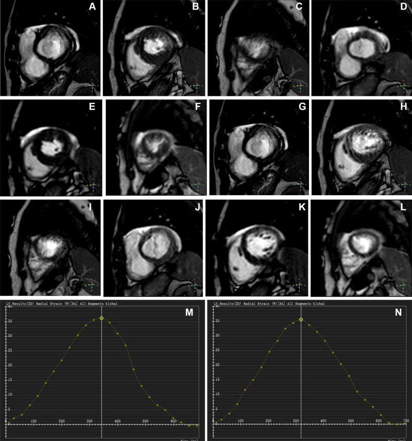
The AI-CS single-shot cine demonstrated significantly better image quality compared to conventional cine, particularly in participants with arrhythmia, with fewer mistrigger events and motion artifacts. Mistriggering can occur when a patient has arrhythmia, causing the scanner to miss the intended phase. Motion artifacts in MRI are common image disturbances that can be caused by physiological (breathing, cardiac) or involuntary movement during data acquisition.
AI-CS also showed good-to-excellent agreement with conventional cine for measurements of biventricular volumes and left ventricular mass. In cases where conventional cine failed, AI-CS provided ejection fraction—the measurement of how much blood the heart’s left ventricular pumps out—comparable to values from echocardiography.
“The AI-CS sequence effectively avoided the cardiac motion artifacts commonly caused by mistriggering in conventional cine,” Zhang said. “It also demonstrated a shorter mean acquisition time while providing improved image quality, particularly in the visualization of the endocardial border, epicardial border, papillary muscles and cardiac motion.”
The success rate for the AI-CS single-shot cine sequence in the study was 100% compared to 88% for the conventional cine sequence, underscoring the challenge of using cardiac MRI to accurately assess ventricular function in patients with arrhythmia.
“The AI-CS framework offers a promising alternative for cardiac MRI examinations in the clinical setting, where long acquisition time remains a major challenge,” Zhang said. “Further optimization of the AI-CS framework, particularly regarding image contrast and artifact reduction, will enhance its applicability in routine clinical practice.”
Source: Radiological Society of North America
27.03.2026