Minimally invasive hip replacement
An anterior approach with the additional support of CT assisted 3-D planning software
By Dr Sebastian Radmer, of the orthopaedics and rheumatic surgery department at Immanuel Hospital, Berlin
Implanting a total hip prosthesis is a major surgical operation that involves severe pain and significant blood loss. Minimally invasive techniques for implanting these prostheses have recently been arousing increased interest. The aim of minimally invasive surgery (MIS) is not primarily to shorten the incision but to lessen tissue damage. It is a question of avoiding structural damage to the muscles, by their being split, bruised or torn, and damage to function, from muscle origins being detached. Blood loss and postoperative pain are supposed to be reduced and as a result the period of rehabilitation is said to be not so long.
However, high standards of safety and the longevity of the implant must not be compromised by reduced intra-operative visualisation. This means precise pre-operative planning to facilitate positioning of the implant with accuracy. Planning is in general performed using templates on conventional X-ray photographs, with factors such as different enlargement, angles of projection that are not always accurate and representation of the total volume clearly reducing precision. The aim of our study was to investigate both the clinical results after implantation of a THP via an anterior minimally invasive approach, and the clinical application of 3-D planning software.
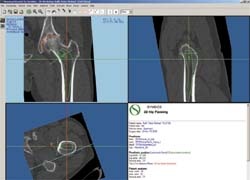
117 consecutive patients attending our clinic (mean age 74.8 (41-83) years) received a total hip prosthesis (Hilock Line cup, Arcad stem, SPS and SPS modular stem, made by Symbios, Yverdon, Switzerland) in a prospective study. All patients underwent spiral CT pre-operatively for 3D planning. The data were processed on an external workstation for 3D planning using special software (SYMBIOS® 3D Hip Plan), and produced an exact view of the acetabulum and femur in all three planes. After establishing the pelvic axis and determining the original centre of rotation, first the acetabular cup was positioned, followed by the stem.
Surgery was performed with the patient supine, and access was via a minimally invasive anterior approach which used the space between the tensor fasciae latae, the gluteus medius and minimus lateral muscles and the sartorius and rectus femoris medial muscles. Specially curved retractor hooks and an angled milling cutter were used during the operation. All the patients were investigated pre-operatively using the Merle d’Aubigné Score and monitored in the same way at 3, 6 and 12 weeks post-operatively, and conventional X-ray checks were also performed post-operatively. Pain was assessed daily up to the 7th post-operative day using the Visual Analogue Scale (VAS).
The surgical technique was able to be performed on all the patients, and the incision length was on average 7.9 cm. Mean operating time was 69 minutes, and mean blood loss in 24 hours was 365 ml. The mean value for pain on the VAS was 7.9 pre-operatively, 2.5, three days post-operatively and 1.4, seven days post-operatively. The mean pre-operative Merle d’Aubigné score was 10.2. Post-operatively it was 15.4 after 3 weeks, 16.9 after six weeks and 17.2 after 12 weeks. The CT plan relating to the cup could be implemented precisely in 108 of the 117 patients (92.3%). In three of the 117 patients, (2.6%), an individual stem was implanted as no modular stem could provide adequately in this respect. The plan was able to be implemented in 101 (88.6%) of the other 114 patients. All the prostheses were implanted without cement, and no stem burst occurred. A total of six complications arose: one prosthesis infection, two wound healing disorders, and three instances of irritation of the lateral femoral cutaneous nerve. No signs of loosening were detected on X-ray.
Implantation of a THP via the minimally invasive anterior approach is a safe procedure that allows the components of the prosthesis to be correctly positioned. It is equally possible to treat muscular patients and obese patients using this type of approach. Blood loss is slight, patients clearly suffer less from post-operative pain, and the duration of the rehabilitation required is considerably reduced. It is particularly useful to be able to combine the minimally invasive approach with a pre-operative 3-D hip plan: pre-operative simulation of various implants and their positioning is thus possible and the centre of rotation of the hip can be optimally determined and reconstructed. Complications that might possibly occur intra-operatively can be identified before surgery is undertaken, and avoided.
E-mail: s.radmer@immanuel.de
30.10.2007