Instead of the “Tube” – An Innovative MRI is open for all Patients
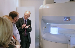
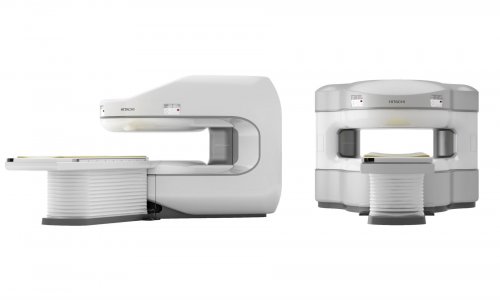
Modern magnetic resonance (MR) tomographs with very strong magnetic fields produce images of steadily increasing precision and are currently gaining more and more importance in radiological diagnostics. In conventional MRI the patient lies in a tunnel, which many people find difficult or even impossible to do, given a scan time of approximately 20 minutes. Physicians of the Joint Radiology Practice at Paderborn’s Medical Center have been working since September 2009 with the Europe-wide unique 1.2-Tesla Highfield MRI OASIS™, manufactured by Hitachi Medical Systems, which features an open architecture instead of a “tube”.


Large medical appliances such as computer tomographs and magnetic resonance tomographs have become indispensable in radiological diagnostics. They are also expected
and asked for by the patients. However, particularly the conventional Mr systems, with their scanning tunnel that measures a mere 60 to maximally 70 centimeters in diameter, are often the cause of great fear. According to the experiences of the radiologists in Paderborn, almost every fourth patient suffers from claustrophobia. As a result, examinations either do not take place, have to be interrupted, or can only proceed under sedation with drugs. Often, images cannot be evaluated because of motion artifacts. Not only in this regard does the OASIS™ represent a great advancement.
The radiologists have already realized their main idea with consequence as early as in 2009, upon drafting the concept of their new offices. The patient is accordingly perceived foremost as a human being who has his own needs, complaints and fears. “Not the patient shall have to adjust himself to the circumstances prevailing on site, instead, we want to make it possible that each patient can be examined in our practice,” explained Dr. med. Carsten Figge, one of the four partners owning the joint medical practice. The same criteria were also crucial to the selection of the new Mr tomograph. The requirement was to find an open system which distinctly enhances patient comfort and concomitantly provides the highest capacity possible along with diagnostic reliability.
Having a field strength of 1.2 Tesla, the OASIS™ is the first high-field MRI in open construction worldwide. The range of vision encompasses 270° and the scan surface inside the tomograph measures 2.5 meters in width. The examination table allowing patients weighing up to 300kg has a range of horizontal movement which exceeds somewhat more than 2 meters. Hence the system is indeed open for all patients. Its technical equipment
assures, among other features, an invariably homogeneous magnetic field and a maximum spatial resolution at shortened imaging times. Owing to its vertically arranged magnetic field, the 1.2 Tesla OASIS™ produces an imaging quality which is comparable with a conventional MrI having a field strength of 1.5 Tesla.
As Dr. med. Jürgen Wiesmann reported, well over 850 scans have been performed with the OASIS™ since mid-September 2009. The desired advantage of the open architecture has already proved its worth: patients had less fear of the MrI instrument. This has been most obviously the case with little children. For example, the practice in Paderborn was able to examine a 4-year-old girl whose arm had been in a plaster cast after a fall.
Thanks to the open architecture the child could not only be put into an optimum position in the center of the magnetic field (isocenter). Sedatives or narcosis were also unnecessary because she had her mother beside her and was able to touch her throughout the entire duration of the scan. The images showed the radiologists a fracture near the elbow which had not been discovered previously.
Elderly patients suffering from pain and limited mobility, obese patients, but also the particularly broad-shouldered basketball players of the premier division team, the Paderborn Baskets, also profited from the Hitachi open MrI system. Due to the combination of open architecture and highest performance Dr. Figge and his colleagues can now dedicate themselves to new important MRI applications, such as interventional MRI abdominal imaging, angiography and mammography.
13.07.2010


