News • Roentgen Professorship 2020
Inspiring young radiologists to take up research
Norfolk and Norwich University Hospital (NNUH) Consultant Radiologist Tom Turmezei has been awarded the prestigious Royal College of Radiologists’ Roentgen Professorship for 2020, a role created to encourage trainee radiologists across the UK to participate in research.
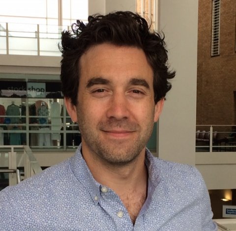
“Research is absolutely key to making discoveries that will improve the diagnosis and treatment of patients in the future,” said Tom. “I’m honoured and delighted to be chosen for this role and, having benefitted from mentorship early in my own career, am passionate about opening young doctors’ eyes to the endless possibilities of research and encouraging them to include it in their careers.”
Roentgen Professors are consultant clinical radiologists with an established research record and are selected because of their ability to inspire others. Tom will visit at least five UK training schemes during his year-long tenure to deliver lectures and tutorials on his research work and inspire others to follow a similar path. “Radiology is such a fast-moving field, using so much rapidly-developing technology, that it’s our responsibility as radiologists to ensure we understand how we can best use it for our patients,” he said.
I understand the importance of giving support and encouragement early on and the difference that meeting inspirational people can make
Tom Turmezei
“Radiology is such a fast-moving field, using so much rapidly-developing technology, that it’s our responsibility as radiologists to ensure we understand how we can best use it for our patients,” he said.
Tom’s research area is musculoskeletal radiology. He is also Radiology Research Lead at NNUH and an Honorary Senior Lecturer at the University of East Anglia. His Wellcome Trust PhD research project at the University of Cambridge led to the development of an algorithm that measures patients’ joints using 3D mapping from routine CT scans and could help detect osteoarthritis earlier, enabling patients to make lifestyle changes and start therapies before their condition becomes debilitating. The technique, called Joint Space Mapping (JSM), is potentially twice as good as X-rays at detecting small structural changes in the bone and could be used to screen people at risk of developing the disease within the next few years. “My work was unusual because I studied for my PhD in the Department of Engineering at Cambridge to develop the Joint Space Mapping technique,” he said. “I’m continuing to analyse new data with the University of Cambridge, the Icelandic Heart Association and Kansas University Medical Center with the aim of implementing this technique in clinical trials.”
Tom has also used his expertise by CT scanning a variety of artefacts, including Egyptian coffins and mummies, artists’ mannequins and cremation remains and is Imaging Editor for Gray’s Anatomy, the 42nd edition due in 2020. “I understand the importance of giving support and encouragement early on and the difference that meeting inspirational people can make,” he said. “Now I’m hoping to inspire others in the way I was.” Tom is the second NNUH consultant to be awarded the Roentgen Professorship, which also means sitting on the Clinical Radiology Academic Committee for two years. Fellow musculoskeletal radiology consultant Andoni Toms held the position in 2011.
Source: Norfolk and Norwich University Hospital (NNUH)
17.07.2019


