Innovations in non-invasive diagnostics
Report: Bettina Döbereiner
Although the 17 lectures delivered at this year‘s Medical Technology Congress in Berlin, Germany, focused on topics ranging from experimental and clinical research
to routine daily diagnostic methods, the pervading interest was in the improvement, development and distribution of non-invasive imaging devices and corresponding software.


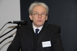
In his welcoming address, Professor Karl Max Einhäupl, Chairperson of the Board of the Charité - University of Medicine Berlin, emphasised: ‘Because of its lower risk of complications the means of non-invasive methods in diagnosing diseases become increasingly important.’ One of these, the liver function test LiMAx, was presented by Johan Lock from the General-, Visceral and Transplantation surgery department at Charité Berlin. This test was invented by Charité Professor Martin Stockmann and developed in close cooperation with physicists at the Freie Universität Berlin.
The measuring method indicates the enzymatic activity and thereby liver function, and it is the first method to provide quantitative and immediate results at the bedside. Aiming to replace the usual blood analysis, the entire procedure takes about 30 to 45 minutes (injection of 13C-methacetin, metabolised only in the liver, and flow-through-breath analysis). Apart from the advantage of a real-time indication, measurement of enzymatic activity in the liver gives more precise information about liver function than the indicators of blood measurement so, for example, liver fibrosis and cirrhosis can be detected at a very early stage of disease. The method also enables a valid calculation of the risk of postoperative liver failure in surgery by preoperative volume planning. Finally, the test enables very close monitoring of liver regeneration. In 2010, the device obtained CE Certification and is in use in all six university hospitals in Germany.
According to Johan Lock, the costs for the measuring device - distributed by Humedics, a spin-off company from Charité and Freie Universität Berlin - actually amount to €50,000. Another promising development was presented by Professor Erwin Keeve, from the Berlin Centre for Mechatronic Medical Technology, a joint Excellence Centre by Fraunhofer Gesellschaft and Charité. With Charité clinicians Professor Norbert Haas, director of the Centre for Musculoskeletal Surgery and Professor Bodo Hofmeister, director of the Cranio-maxillofacial Surgery department, and the company Ziehm Imaging Ltd, he and the team are developing an intraoperative X-ray imaging system that will provide a 3-D image recording method without surrounding the patient, allowing simultaneous intra-operative use of imaging. Three dimensional imaging, while measuring less than 360 degree angle, is mathematically impossible, but the team calculated and defined the specific error function and thus could minimise and allocate the errors in imaging. ‘Only the diagnose-ability has to be ensured, therefore image quality and pixel resolution can be ignored to a great extent,’ Prof. Keeve explained.
In 2010, their Orbit project receive the innovation prize of the Federal Ministry of Education and Science, and Prof. Erwin Keeve is optimistic that he might present the first prototype in five years and finished product in seven or eight years. Regarding the general development of new technologies, Prof. Einhäupl proclaimed in his keynote speech: ‘We need a new culture of industrial cooperation that allows us to jointly develop new products – but also foresees a fair sharing of the intellectual property rights and the benefits.’ The health industry takes another line in this regard, he pointed out, but he is convinced that the industry could and should be persuaded by fair sharing models, in a win-win situation for both partners in the future.
* The event was again initiated and organised by TSB medici, the medical technology division of the TSB Innovation Agency Berlin Ltd. in cooperation with the Charité Medical University and the city‘s chamber of commerce and industry.
20.06.2011


