Imaging could improve treatment of COPD
Magnetic resonance imaging (MRI) and computed tomography (CT) provide important information on the symptoms and exercise capabilities of people with mild-to-moderate chronic obstructive pulmonary disease (COPD), according to a new study published online in the journal Radiology. Researchers said the findings point the way to better treatment for some COPD patients.
COPD is a progressive disease of the lungs that affects approximately 65 million people worldwide, according to the World Health Organization. Diagnosis often involves spirometry, a lung function test in which the patient takes a breath and exhales forcefully into a tube connected to a machine. This test, which produces a figure called the forced expiratory volume in one second (FEV1), has limitations.
"COPD is a very heterogeneous disease," said study co-author Dr. Grace Parraga from the Robarts Research Institute in London, Ontario, Canada. "Patients are classified based on spirometry, but patients with the same air flow may have different symptoms and significant variation in how much regular activity they can perform, such as walking to their car or up the stairs in their home."
FEV1 does not necessarily reflect the whole picture of the lungs, but airways disease and emphysema can be directly measured with imaging. Parraga and colleagues set out to develop a way to explain COPD symptoms and exercise capability.
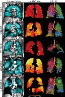
For the study, they performed conventional CT and inhaled noble gas MRI, a technique for visualizing air spaces in the lungs, on 116 people with COPD, including 80 with milder disease. The patients also underwent lung capacity testing, filled out a quality of life questionnaire and took a six-minute walk to measure their exercise tolerance over a short period of time.
The results showed that in mild-to-moderate COPD patients with modestly abnormal FEV1, MRI measurements of emphysema were strongly correlated with exercise limitation, while both CT and MRI measurements of emphysema helped explain symptoms.
The implications of the findings are significant for patients with mild COPD and abnormal FEV1, according to Parraga.
"FEV1 doesn't tell the whole story," she said. "With lung imaging, we can look at patients with mild disease much more carefully and change treatment if necessary."
Findings associated with COPD include emphysema, or damage to the air sacs in the lungs that prevents people from getting the oxygen they need, especially during exercise. While no cure exists for emphysema, there are steps people can take to mitigate symptoms. Emphysema is under-recognized as a source of COPD, Parraga said, and as a result, patients may be getting suboptimal treatment.
"One in four hospital beds in Canada is occupied by a COPD patient, and many of them return to a hospital because they're not being optimally treated," Parraga said. "Our study shows that when COPD symptoms and exercise limitations are discordant with FEV1 measurements, we should consider using lung imaging to provide a deeper understanding of the patient's disease and to help improve their quality of life."
The researchers plan future studies to see if imaging can help explain symptoms and disease control in people with asthma, another common lung disease, and cystic fibrosis, a hereditary disorder that damages the lungs and digestive system.
Source: Radiological Society of North America
07.07.2015