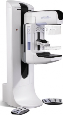
Hologic brings tomosynthesis to breast cancer screening
In May, 2013, the U.S. Food and Drug Administration (FDA) finally took the training wheels off tomosynthesis by approving the use of Hologic's new C-View 2D imaging in place of conventional 2D mammograms previously required as part of a breast tomosynthesis screening exam.

The evidence was compelling. Radiologists found 30% more cancers in first round breast cancer screenings.
To meet the man who played a key role in providing the evidence, plan on attending the presentation of findings from the now-famous Oslo study by Per Skaane, M.D., Ph.D. of Oslo University Hospital Ullevaal.
The study was based on 12,631 screening examinations in a large hospital in Norway.
The FDA approved protocol, called the combo-view, was integrated as a separate arm in the randomized Oslo study where independent readers interpreted both the 2D C-Views and consulted the 3D data set of slices.
Comparing an arm of the Oslo study that interpreted traditional 2D full field digital mammo exams with the combo-view arm, Skaane reported the stunning increase in the cancer detection of 30% with digital breast tomosynthesis (DBT).
"This means we have a new test that increases specificity and with a superior visibility and conspicuity for breast cancer, which increases sensitivity, the most important thing in my view," he said.
The mean interpretation time in the batch reading mode was 45 seconds for traditional 2D mammography exams and 91 seconds in the combo-view mode, he said.
"Yes, it is a doubling of the reading time," Skaane said. "But if we find significantly more cancers, doesn't this justify 45 seconds longer? That is the crucial question."
He and his colleagues in Oslo decided that doubling of reading time is acceptable in the screening setting.
Time spent reading exams is the critical factor required to win adoption in the high-patient throughput setting of breast cancer screening, and reading multiple slices of a 3D DBT exam simply takes too long.
Here Hologic made a strategic shift by dropping an insistence on the quality of the 3D reading to produce 2D images more familiar to mammographers and that fit in their fast-moving workflow.
Flattening the 3D image set to 2D is much more sophisticated than stacking multiple slice views. There is complex modelling and processing behind these synthetic 2D images.
Applying a capability for image analytics and automated detection it acquired with the purchase of R2 Technologies, Hologic introduced C-View, a 2D synthesis of images taken from the 3D dataset that are presented in the familiar mediolateral oblique (MLO) and cranio-caudal (CC) views.
"I think most of us will agree the image is nicer," Skaane said in presenting side-by-side examples of traditional mammogram and a C-View 2D image in October to French radiologists
"The important question is whether in combination with tomo we get the information from the synthetic 2D that we need," he said, adding that the dose is approximately the same as with conventional 2D mammo.
"If we compare the pure 2D with the combo-mode [C-View + tomo] there is no increase in detection rates for DCIS [ductal carcinoma in situ]," he noted
"What we pick up using tomosynthesis are invasive cancers presented as spiculated masses and distortions. This is the main benefit of using Tomo," he concluded.
02.12.2013
- breast cancer (626)
- cancer (1026)
- economy (1046)
- mammography (258)
- radiology (730)
- research (3487)
- screening (218)


