High speed MRI devices gain use in foetal evaluation
Cynthia E Keen reports
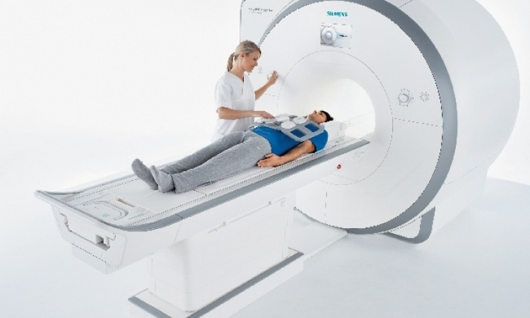
Magnetic resonance imaging is gaining increasing importance as a second imaging process in prenatal diagnosis in addition to ultrasound examination, according to Dr Daniela Prayer, a paediatric radiologist at the University Clinic for Radiological Diagnostics at Vienna University Hospital.
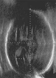
week 25+4, sent to MRI
for screening for brain abnormalities after ultrasound examination showing slight asymmetric ventriculomegaly
Speaking at a press conference at the ECR, she said that, used in conjunction with 3-D ultrasound and Doppler or colour-Doppler ultrasound, MRI could establish itself as the means by which gynaecological, gastrointestinal, urological and possibly oncological processes in foetuses with potential abnormalities could be imaged. MRI has historically been used when ultrasound examinations were impaired by a lack of amniotic fluid, unfavourable foetus position and/or excessive obesity of the mother. Both ultrasound and MRI are non-invasive, non-radiation emitting diagnostic procedures.
Use of high speed sequences in MRI now enables radiologists to obtain interference-free imaging. Almost all foetal body parts, such as the face, neck, thorax and abdominal organs, as well as the mother’s tissue can be reliably and precisely visualized. Brain development can be assessed with almost histological precision. Surplus kidneys can be detected and renal tissue can be superbly visualized. It is also possible to detect intrinsic movements of organs in real time. ‘These developments make precise and detailed diagnoses easier,’ Dr Prayer explained.
Vienna University Hospital has been using MRI for prenatal diagnostics since 1998. When a foetal abnormality is suspected in an ultrasound examination, MRI is used as an effective additional examination method. About 50% of pregnant women whose foetuses are suspected of having irregularities in the central nervous system and lungs have an MRI performed at Vienna University Hospital. Dr Prayer said that, in Austria, the cost to perform a specialized 3-D ultrasound procedure and an MRI are comparable. ‘In comparison with older MRI systems, the high speed systems allow the visualization of moving processes,’ she pointed out. ‘If a foetus with a stenosis in its oesophagus has problems swallowing amniotic fluid, this can be detected in the gradient echo sequence. This enables obstetricians to plan for possible post-birth surgical procedures.’
Dr Prayer said that the possible risks of an MRI procedure include the warming of the mother and foetus and the fact that the examination may cause stress to both the mother and the foetus. The foetus may also be exposed to vibrations and noise. There is no indication that this causes harm, but this fact has not been ruled out either.
‘We believe that the benefit to the mother and her developing foetus is far greater from a clinical perspective than the risks we can identify,’ she concluded.
03.06.2008