High-res cardiac images available at peak stress
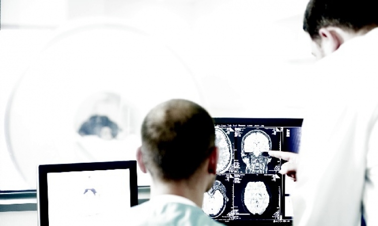
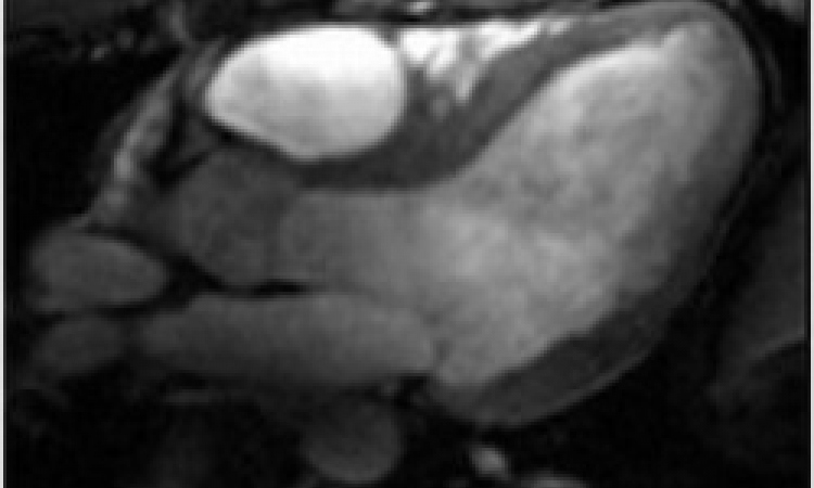
While treadmill exercise stress testing is essential to detect cardiovascular disease, gaining clear cardiac images at peak stress level are not easy to gain using standard testing procedures. Now, however, researchers at the Ohio State University Medical Centre have designed equipment to provide high-resolution cardiac images at a critical testing stage, with results in under one hour.
‘In the past, we were constrained by the time lapse between the completion of exercise and capturing the images,’ explained Orlando Simonetti PhD, associate professor of internal medicine and radiology. ‘We now have the ability to exercise patients to peak stress and obtain a high definition image of their heart within 60 seconds, which helps us more accurately identify exercise-induced abnormalities. OSU Medical Centre is the only place in the world performing treadmill exercise stress tests inside the MRI scan room.’
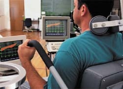
The standard design of treadmills has made exercise stress testing a challenge near the large magnetic field generated by the MRI equipment. Simonetti and team, working with graduate students from Ohio State’s College of Engineering and faculty from the OSU Agricultural Technical Institute, modified a treadmill for use in close proximity to the MRI examination table. Magnetic components were replaced with non-magnetic stainless steel and aluminium equivalents.
While patients perform the treadmill exercise test, they are monitored using a 12-lead electrocardiogram system, which is disconnected after exercise. Heart rate and rhythm are then monitored with a wireless, MRI-compatible electrode unit while patients undergo a rapid, real-time imaging procedure that takes less than a minute.
Clinicians are excited about the possibilities. ‘While current forms of stress testing have been helpful, combining exercise stress with cardiac magnetic resonance imaging allows us to better measure the presence and extent of heart disease with a clarity not previously possible,’ said Dr Subha Raman, associate professor of internal medicine in OSU Medical Centre’s cardiovascular medicine division.
01.09.2008