Enhanced image quality and improved workflow
At this year's RSNA, Philips not only showcased their latest technology, the Achieva 3.0T TX MRI scanner, but also provided insights in their current activities to improve workflow in the radiology department: in collaboration with the University of Chicago Medical Center the company conducted a closed loop imaging research trial.
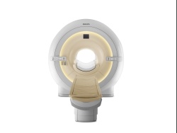
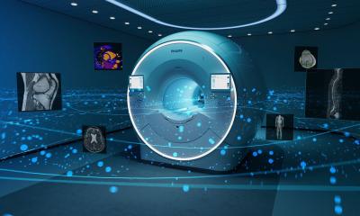
MRI scanner with patient-adaptive technology
The Achieva 3.0T TX enhances image quality, provides up to 40 percent greater scanning speed and helps ensure fewer retakes through increased image uniformity. Capturing the correct information on the first scan in a shorter amount of time delivers more convenience to the patient and increases patient throughput potential for healthcare providers. Additionally the new scanner provides consistent results across a broad range of clinical applications and patient sizes.
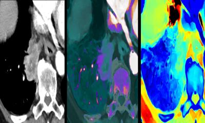
In the past, 3.0T imaging has been challenging for certain clinical procedures due to dielectric shading effects and local specific absorption rates (SAR). Philips’ new, proprietary, MultiTransmit technology addresses these issues at the source with multiple RF transmission signals that automatically adapt to each patient’s unique anatomy. As a result, the new Philips MRI scanner delivers superb diagnostic images for even the most demanding high field applications such as breast and liver imaging.
Reducing errors and improving quality of care
At RSNA, Philips announced that it is partnering with the University of Chicago Medical Center (UCMC) on a first-of-kind closed loop imaging research trial. The purpose of the ongoing trial is to take a holistic view of radiology workflow and integrate information systems together with imaging systems to remove inefficiency at all points within the imaging loop. The so-called “imaging loop” clock starts when the physician decides to order an imaging exam and ends when the results of the test are communicated to the doctor and eventually the patient.
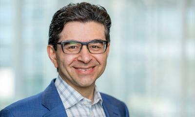
Led by Paul J. Chang, M.D., Medical director, Enterprise Imaging for University of Chicago Hospitals, UCMC has initiated this iterative study with the goal of reducing errors and improving quality of care and patient outcomes. Together with Philips, UCMC is integrating the technologies used throughout the radiology suite so they “talk” to each other across a smart, automated and open platform, seeking to streamline imaging procedure steps to save minutes, not seconds, during the entire imaging loop. Helping to improve efficiency of radiology workflow may help UCMC reduce patient waiting time, repeat scans and the time needed for appointments.
“To stay relevant in healthcare, we simply can’t keep using the same workflow model,” said Dr. Chang. “Radiologists need to provide the highest quality diagnostic results to the patient’s physician while obtaining the best images on the first scan, every time, without compromising patient care. This requires an orchestrated workflow that can only be achieved if all systems used in the imaging loop are integrated and provide an interface allowing information to flow seamlessly from one step to the next, minimizing inefficient and potentially distracting busy work for the radiologists and technicians.”
As a first necessary step of the research trial process, Philips and UCMC have successfully identified major bottlenecks and non-essential steps in the imaging loop, and have determined the right technologies to help solve the inefficiencies facing radiologists and technologists. During this initial research phase, UCMC replaced the current paper-based CT protocol system ─ manual entry of a list of images requested, as well as contrast and dose requirements ─ with an automated electronic patient protocol system which uses a Philips tablet PC for wireless access to relevant patient and scanning protocol information. Protocol settings are automatically communicated to the CT scanner, simplifying CT scanning by sending all relevant clinical information and protocol definition without requiring manual entry. Event-driven alerts provide updates throughout the entire imaging process and notify radiology staff when images are ready for processing. Overall, the radiology department was able to eliminate non-essential work and paper trails. Upcoming iterations of this project will seek to incorporate additional, customized solutions that directly address the bottlenecks identified in the imaging process.
04.12.2008