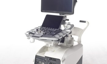
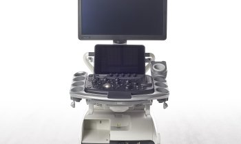
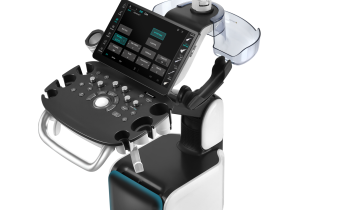
Elastography - Hitachi expands technology applications
Compelling evidence validates tissue strain exams in new medical fields. John Brosky reports
In recent years Hitachi Medical, the pioneer in developing ultrasound elastography to diagnose tissue stiffness, has seen competing systems launched that claim to have the same capability. ‘We are approaching a point where elastography will be considered a standard feature, call it the E-mode, for ultrasound,’ Heinz Schreiber, head of Ultrasound for Hitachi Medical Systems Europe told European Hospital.
Yet Hitachi, which introduced this revolutionary technology in 2004, continues to lead a field of ‘me-too’ products by offering compelling clinical evidence of the effectiveness of elastography examinations and by expanding the applications for this new science.
‘Elastography is fast becoming like air conditioning in a car where it is difficult to find a new car without it,’ he said. However, Hitachi is the only ultrasound maker offering elastography on all of its scanners, Dr. Schreiber pointed out, adding that competing elastography systems offer this valued feature only on more expensive, high-performing ultrasound platforms.
Hitachi Real-Time Elastography (HI-RTE) is also compatible with over 20 different transducers.
Well-established as a valid technique for more precise examinations of suspect nodules in the breast, thanks to Hitachi-sponsored published studies, new evidence from the company shows that elastography can be used as well for diagnostics of musculoskeletal disorders, the thyroid gland and lymph nodes, the prostate and testicles, as well as in dermatology to assess melanomas.
Dr Mercedes Roca, at the University of Zaragoza, Spain, concluded that HI-RTE is a promising method for the quantification of the elasticity of musculotendinous elements in sport trauma.
Dr Ferdinand Frauscher, at the University Hospital of Innsbruck, Austria, found advances in Hitachi elastography improved prostate cancer diagnosis and that subsequent biopsies of areas targeted by HI-RTE were three times more likely to detect cancer than conventional systematic sampling.
Clinical experiences from Hitachi indicate the technology is also useful in obstetrics to measure the elasticity of the upper walls of the uterus to determine a risk of early delivery.
Corrine Bricout, Manager for HI-RTE in France, recently demonstrated the capability for clinicians at the recent French radiology congress. ‘For the clinician,’ she said, ‘the clinical utility of elastography is continually advancing. There is greater and greater validation for diagnoses using elastography for muscle tears, for example the Achilles tendon.’
Elastography allows a radiologist to say with greater confidence that a condition is of concern or not, to give a same-day, real-time diagnosis. ‘We have already seen there is a tremendous psychological benefits of this for a woman with breast examinations, as she no longer needs to live with uncertainty about a nodule for six months to know if her doctor’s concern is justified or not.’
This same insight and precision can be of great benefit for other patients, Corrine Bricout added, as well as giving radiologists greater confidence in their diagnoses.
Hitachi measures of strain rate offer a real-time relative quantification of the scanned tissue for greater objectivity in assessing the elastography image.
This HI-RTE scoring of tissue stiffness is also available across the range of Hitachi ultrasound scanners.
Bricout demonstrated how the measures of a signal received from suspected tissues is compared to a reading of similar tissue from the same organ that is known to be healthy. A simple example, she said, is for an operator to compare the scan of healthy tissue in the left breast with a suspected nodule in the right breast.
Continuing to build its lead as the pioneer in elastography, Hitachi is currently conducting clinical evaluations for a quantitative algorithm for liver fibrosis. ‘The liver is the most exposed organ of the body because it is a chemical factory through which your entire metabolism passes,’ said Dr Schreiber. ‘The degree to which there is fibrosis in a liver becomes vitally important for the health of a patient. We have developed nine parameters to represent a spectral histogram that quantifies the degree of fibrosis of the liver,’ he said. ‘This is a very complex diagnosis, and the Hitachi capability is comprehensive providing in a non-invasive examination the kind of information essential to a diagnosis that today can only be achieved with surgical biopsies.’
While elastography is the measure of elasticity in the surrounding tissue, it is not precisely tissue characterisation, he explained, where the quantificative results of the enhanced capability for abdominal examinations begins to approach true tissue characterisation for the liver and pancreas.
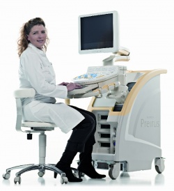
The new technology will be onboard the new Hitachi Preirus compact model, planned for introduction at the European Radiology Congress in Vienna in March, 2010.
‘There is an enormous market in gastroenterology and we expect to have an advantage of several years in the clinics,’ he said, because competitors will need to conduct university level research to set their own parameters for the quantification by ultrasound of these organs.
Details: www.hitachi-medical-systems.eu
18.11.2009


