Dual-Source CT
Aiming for even higher performance levels
X-ray computed tomography (CT) has shown an absolutely remarkable and impressive increase in its performance characteristics for many years - remarkable because the modality was declared dead in the 1980s, impressive because these developments seemed impossible to many, for technical and for physics reasons.
Today, effective scan times per image of well below 500 ms and isotropic spatial resolution below 0.5 mm are a clinical reality. All fields of application profit from this performance, especially cardiac imaging and CT coronary angiography, which rely on it.
The renaissance of CT began, in the early 1990s, with the introduction of spiral CT, allowing fast and continuous scanning of complete organs and anatomic sections. To make it a practically usable reality the development and introduction of array detector technology and of more powerful X-ray equipment was necessary. The first multi-row detectors became available in 1998. After mastering the technology, manufacturers were quick to add more and more rows and succeeded in making multi-row CT affordable for a wide user spectrum. This was the start of the ‘slice race’. At present, multi-row CT technology has become standard and the simultaneous acquisition of 64 slices constitutes the state of the art.
Effective handling and evaluation of large thin-slice data sets, with up to 4000 slices per patient, has turned into a key challenge for the CT industry. Adding more rows to the detector array, and thereby providing possible acquisition of more slices simultaneously, is an option that could be made available easily. It is still open as to drawbacks associated with the use of wider detector arrays, or if related data management challenges can be solved, and what costs might be expected - and finally whether there is clinical demand for it.
Increasing the X-ray power necessary for faster and faster scanning is more of a problem than increasing the number of detector rows. To provide the number of quanta necessary for a specified level of image quality demands a certain effective mAs product. Every time we reduce the rotation time of the scanner, the tube current or X-ray power must be increased accordingly, to keep the mAs product constant and to maintain the signal to noise ratio. Manufacturers have responded to these demands; these days, 80 to 100 kW X-ray units are on board the rotating gantries. However, just as in electron beam CT, a concept that, meanwhile, was given up, the maximum tube currents are limited not only because of limited generator power, but because of anode durability. The peak power levels that are necessary for typically 5 to 20 seconds cannot be increased easily.
Why should we ask for shorter rotation times and higher X-ray power levels after all? CT seems to master almost all demands imposed by clinicians. This may seem true when looking at the impressive and partly spectacular results presented by researchers, clinical radiologists and the industry, preferably rendered in colour 3-D and animated fashion. However, it is known that some demands have not been met fully yet. CT coronary angiography, one of the most demanding applications, yields perfect results in many cases, but not in all. Typically 10% of clinical investigations remain inconclusive due to motion non-sharpness. Just the same, X-ray power may prove insufficient in obese patients, causing delays in the examination. And, after all, there is always the request for more specific information than provided by the standard CT values.
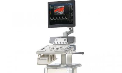
Dual-source CT (DSCT) appears to be one of the potential solutions for increasing CT performance levels further, without technological overkill. It basically consists of a scanner that features two X-ray sources and two detectors. Respective ideas were promoted in the1970s, when CT scanners were still too slow for many applications. Manufacturers managed to provide improvements and requests for respective innovations died down, particularly since the focus was put on MR imaging. Above all, it was cardiac imaging that made respective technical approaches attractive again for CT. Meanwhile the world’s first dual-source CT system, a Siemens Somatom Definition, has been installed at the Institute of Medical Physics (IMP) in Erlangen and has been under clinical evaluation since October 2005. This features two 80-kW X-ray systems and two 64-slice acquisition systems under one cover. Patients and users do not notice much difference, since the system’s outer dimensions have not changed significantly. Efficient engineering increased the components packing density and provides the increased performance within a standard CT gantry. An in-depth analysis of the considerations presented above and the technical possibilities was recently given by this author in the textbook Computed Tomography (Publicis Corporate Publishing, Erlangen 2005).
The results that dual-source CT provides are impressive and a particular pleasure for any CT aficionado. One of the primary goals - high reliability and robustness in cardiac imaging - has been achieved to a high degree. Effective scan times can be guaranteed to be 83 ms as a maximum; with respective multi-phase retrospective cardiac interpolation algorithms these can be lowered to 50 ms and less, as introduced by [T Flohr et al, European Radiology, February 2006]. A first clinical publication by Stephan Achenbach et al. [European Journal of Radiology, in press] reported that all coronary arteries were imaged successfully with 98% of the examined coronary artery segments free of motion artefacts.
CT seems to have made available another impressive step in performance level. It is, above all, the increase in scan speed, which was always one of CT’s major advantages. But it may also provide improvements in diagnostics specificity. Combination imaging with PET/CT has already brought major advances in this direction. Dual-energy imaging with dual-source CT may provide further advances and will be, in years to come, a hot topic in CT research.
My conclusion: CT is alive!
Contact: willi.kalender@imp.uni-erlangen.de
01.03.2006