Article • Advancing kidney disease investigation
Digital spatial profiling: new ways for diagnostic histopathology
Digital spatial profiling (DSP) is emerging as a powerful technology in helping specialists investigate complex kidney disease, according to a leading expert. Professor Renate Kain believes spatial profiling adds significantly to systems biology approaches that will transform diagnostic histopathology. However, she warns that the adoption and effective utilization of the technique is critically dependent on pre-analytics of the samples investigated.
By Mark Nicholls

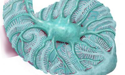
Image source: MedUni Wien
At the Digital Pathology & AI Congress in London, Kain outlined how the technology has applications in immunological disease, particularly for patients with vasculitis, where long-term survival is poor. In her presentation, she gave an overview of DSP’s advantages and limitations, while detailing on selection bias and regions of interest (RoI), pre-analytics, and applications in vasculitis research.
DSP, a technology that analyses gene or protein expression in tissue architecture, is increasingly used to interrogate transcriptomic profiles in tissue sections. A method to investigate specific disease pathways and molecular signatures, it is now providing a technology for use in complex auto-immune diseases of the kidney, said Kain, who is Head of the Department of Pathology of the Medical University of Vienna, Austria, and a histopathologist with a special focus on inflammatory disorders of the kidney.
She said DSP is particularly beneficial with the significant rise in slides produced due to the evolution of an increasing number of biomarkers and next generation sequencing. ‘We have been waiting for spatial profiling technology for pathology for a long time,’ she said.
Combining AI with transcriptomic signatures
The technology is enabling specialists to see clustering patterns and areas of tissue that are most severely affected by disease, the expert pointed out. ‘We can also differentiate and visualise differences between different diseases. With biopsies, we sometimes did not see changes, but what we see here are normal and abnormal changes with a transcriptomic signature that will tell us that there is a disease process ongoing. This is where AI comes in. Several groups are now using the technology and also bringing it into the diagnostic work process.’
Recommended article
Article • In-depth
Focus on digital pathology
Digital pathology opens up a whole new world of possibilities in diagnosis, prognosis, and prediction of diseases. Keep up-to-date with the latest research news, medical applications, and background information on digital pathology.
Kain said different transcriptomic signatures had been found from information contained on a simple slide that previously would have taken many investigations to discover. This has benefits in kidney disease, which she said was a growing worldwide problem. She specifically referred to focal necrotizing glomerulonephritis (FNGN), a rare kidney disease that causes destruction of the glomeruli, the clusters of blood vessels in the kidneys that filter waste from the blood.
Gene profiling using DSP allows investigation of individual glomeruli and to identify distinct patterns associated with different glomerular lesions. That includes expression profiling from small and spatially circumscribed regions of tissue and expression signatures from individual glomeruli with defined morphological characteristic. ‘We can identify single glomeruli and compare predictions from that to see trends, all from a single slide and in spatial context,’ she said.
The importance of proper pre-analytics
We might not need immunohistochemical stains anymore, but spatial profiling and single cell sequencing are methods I trust will change the way we do diagnostic histopathology
Renate Kain
However, Kain underlined the importance of the pre-analytical process and noted limitations of DSP such as throughput sensitivity and limited number of samples per slide. But she said the Nanostring GeoMx DSP platform is suitable to investigate individual glomeruli and identify genes differently expressed between different types of lesions and different disease aetiology.
The expert concluded: ‘Spatial profiling is a powerful tool to investigate disease mechanisms and pathways in tissue at local level across the human disease spectrum. Sooner or later, it will find its way into the diagnostic process, similar to multiplex immune-imaging and proteomics. We might not need immunohistochemical stains anymore, but spatial profiling and single cell sequencing are methods I trust will change the way we do diagnostic histopathology, but with the caveat and prerequisite that the quality of data we analyse are critically depending on pre-analytics.’
Profile:
Professor Renate Kain is Head of Pathology at the Department of Pathology of the Medical University of Vienna with a special focus on inflammatory disorders of the kidney and leads a research group investigating molecular mechanisms of autoimmunity. She is also a partner in the IMI2 project Bigpicture that has been designed to establish a large database of pathology images to accelerate the development of artificial intelligence in medicine.
31.03.2026