Biograph mCT
Signalling the beginning of molecular CT
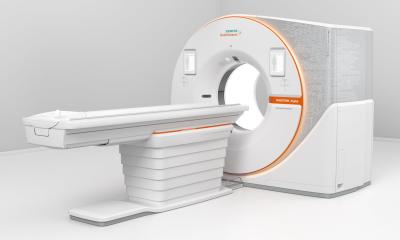
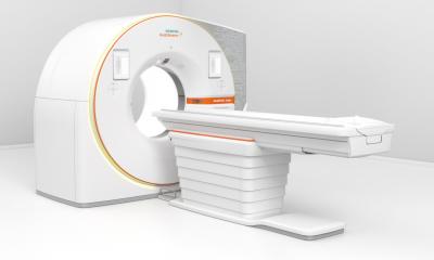
At Medica 2008, Siemens introduced its latest generation PET-CT, the Biograph mCT. During a European Hospital interview, Markus Lusser (ML), worldwide Head of Distribution and Marketing for Molecular Imaging at Siemens, outlined the advantages of the new hybrid system, which aims to extend the spectrum of medical imaging to 'molecular computed tomography'

What makes the mCT different from other PET-CT systems?
Markus Lusser: The Biograph mCT combines our highest-performance CT platform with our latest PET scanner. This means the system combines two high quality modalities in one machine, whereas with conventional PET-CT systems the CT component has limitations for complicated CT examinations. To increase patient comfort, we have now integrated both components in a very small system, so that the scanner has a tunnel length of only one metre. This means that examinations with the Biograph mCT can be carried out in just five to six minutes, whereas conventional machines still take about half an hour.
Does this mean the system is not really a PET but a CT with integrated PET module?
Yes, a powerful CT with high performance PET. With its adaptive dose shield, a scanning area of up to two metres and a maximum time resolution of up to 150 milliseconds – with 128 slices per rotation – the CT scanner ensures pin-sharp and clear images. This makes it suitable for routine diagnostics as well as complex examinations in cardiology and oncology.
These are different medical disciplines. The mCT constitutes the interface between radiology and nuclear medicine. Will these two disciplines be forced into increased cooperation due to this technical development?
I wouldn’t use the word forced, because PET-CT is already an established modality. It means that nuclear medicine specialists and radiologists jointly establish the diagnosis. A specialist in nuclear medicine typically cannot evaluate and diagnose the results of a CT examination, and a radiologist cannot evaluate PET results, so we are supporting closer cooperation with our mCT.
But this has a significant bias towards radiology. Some hospitals in Germany have nuclear medicine departments and professorships. To use the new mCT effectively, radiology and nuclear medicine will now have to work together in one department.
As the manufacturer, we don’t think so much in terms of the structural categories but look at issues from a clinical point of view. Moving away from the situation in Germany, internationally there is a strong trend towards integrated imaging – and we aren’t interested in promoting one group to the detriment of another. Hybrid systems, such as molecular CT, need the expertise of both groups if the potential of ‘molecular imaging’ is to be fully exhausted. For example, this type of CT can actually be used for organ perfusion. However, that requires a radiologist’s expertise in the necessary technological skills. If you then superimpose the result with that of a PET examination – and this is where nuclear medicine comes into play – you gain a third type of measurement, at no extra expense, which can be relevant for fast and precise diagnosis. But the crucial point is that about 95% of all PET-CET users do not use CT for advanced examination protocols, such as contrast-CT.
So they are only driving your luxury limousine in first gear?
ML: Only in first gear, and to pop to the shops, so to speak. And this was why Siemens focused on how to motivate users to make full use of the potentials of a CT examination. The solution: an enhanced CT that can also be used to carry out CT angiography. The mCT can give a surgeon a complete image of the lungs, for example. This clearly shows where the bronchial tubes and tumours are. It can even show blood vessels – and all this in an integrated examination for which the Biograph Molecular CT only requires a fraction of the time that currently used machines take to do it.
20.12.2008


