World Congress of Cardiology 2006
Massive gathering of cardiologists promises vital knowledge exchange
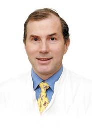
Professor Raimung Erbel, of the West GErman Heart Centre Esse, Duisburg University Hospital aobut the hot topics of this years´congress.

Raimund Erbel
Thus the event promises more than a lively exchange of scientific research results and innovative cardiology solutions. Professor Raimund Erbel, of the West German Heart Centre Essen, Duisberg University Hospital, Germany, forecasts and discusses the likely ‘hot’ topics.
‘Imaging procedures will be a main focus at the World Congress. Currently, 64-slice and 128-slice CT are very topical – and now we are also expecting images from a 256-slice. It remains to be seen how accurate and detailed images produced by the 256-slice are and whether we will gain any diagnostic benefits from them.’
‘Imaging via MRI is also becoming more important in cardiology. Developments in this area are also rapid - some manufacturers have already moved from Tesla 3 to Tesla 7.’
‘There are also some very new developments in radiographic techniques for heart catheterisations. At the University Hospital in Duisburg-Essen we were the first in Europe to install a procedure that combines conventional radiographic technology with ultrasound scanning technology. This brings us a big step closer to the modular development of a catheter workstation. We will present our first experiences and evaluations at the Congress.’
‘We have been closely involved in this development. For years, we had been asking for the development of modular systems for cath labs. Now we have finally succeeded in integrating not only X-ray diagnostics into the cath lab, but also ultrasound diagnostics. The difficulty with this combination has long been the correct ‘match’ between the two diagnostic systems – the fusion of two types of images in a way that ensures a meaningful overall image for diagnosis.’
‘The first system of this kind results from GE Healthcare partnering the Volcano Corporation (see box). The X-ray and ultrasound diagnostics combination gives a cardiologist inimitably
clear images of the coronary and peripheral vascular morphology. The images achieved by merging data from X-rays and ultrasound scans significantly ease the evaluation of the severity of cardiovascular diseases.’
‘This puts cardiologists in a position where they can make decisions on the appropriate therapy options much faster - and safer. This new state-of-the-art technology enables us to shine light onto, so far, dark areas surrounding the causes and progression of coronary and peripheral arterial diseases. Other medical companies also will aim to achieve this type of combination. The next step for cardiology will now be to equip catheter systems in such a way that the X-ray images produced can be directly aligned with CT or MRI images.’
‘This calls for intelligent IT solutions. We are currently working on a solution based on the upgrading of a PACS system, and which enables the combination of different imaging systems at the catheter workstation. There has been a lengthy demand for this combination, but the technical realisation, up to now, has been too difficult. The PACS itself offered no solution here, because the programme was only designed around data and image archiving. However, this type of archiving, without analysis of results, is of little use for diagnosis.’
‘The Hospital Information System (HIS) that we developed brings together all patient data in one place - doctor’s letters, Echo and ECG, X-rays and other diagnostic images. This grouping ensures easy availability of relevant information and facilitates the best possible diagnosis. We developed this system in co-operation with an American company; it is already on the market.’
Asked whether imaging procedures, previously the realm of radiologists, are increasingly entering the realm of the cardiologist, and whether this causes conflict, Professor Erbel said a similar situation occurred in the past: ‘Heart catheterisation used to be the responsibility of radiologists, and, for example in Sweden, this is still the case. However, the worldwide trend is towards an exchange between different medical fields, which then results in new specialisations. So, from the beginning there must be a close co-operation between radiology and cardiology, with the objective of finding synergies in diagnostics.’
‘Take an example from our university hospital. One of our MRI scanners is based at the heart centre and is used jointly by the cardiology and radiology departments. Basically, the cardiologist asks for the MRI images and the radiologist produces them. Both specialists then carry out the evaluation and diagnosis, together. The aim of this procedure is for the clinician who knows the patient to communicate closely with the radiologist to achieve comprehensive diagnostic findings.’
Asked about intervention as another big issue in cardiology, Professor Erbel said: ‘New developments in this area focus on heart valves, i.e. heart valve replacements and reconstructions with the help of catheter technology. This is definitely an important topic in view of the ageing population worldwide.’
‘In the area of drug eluting stents, research is concentrated around finding materials that can form vascular surfaces quicker and more complete and resolve afterwards. We are trying to get away from the type of stent that remains in the body as a foreign object. Two developments in this field look promising – magnesium-stents and those made from polymer plastics. ‘The prevention of cardiovascular diseases will also be among the congress topics. Over the last few years, effective therapies have been developed for lowering harmful low density lipoprotein (LDL) levels, so the research is now focusing on ways to utilise the protective characteristics of high density lipoproteins (HDL). The results of an American study into the subject of progression and regression under the increase of the HDL (i.e. into establishing whether an artificial increase in HDL leads to a regression or progression of the disease) are due to be published this autumn.’
‘This is done with the help of a cholesterol transport system inhibitor. The result of this study could give a real boost to preventive therapy. So, overall we can look forward to some important developments and innovations at the congress.’
On the wall in his office is a certificate stating that he is ‘Man of the year’. Asked about this, the professor explained that it is science related, and awarded by the American Biographical Institute. Prompted further, he added: ‘I was selected to receive the Man of the Year award because of my numerous publications.’
Further details: www.escardio.org/congresses/World_Congress_Cardiology_2006/WCC_2006/
30.08.2006
More on the subject:


