Picture: Carmela de Marco / ETH Zurich
News • Microstents vs foetal urethral strictures
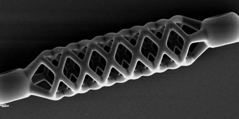
The world’s smallest stent
Researchers at ETH Zurich have developed a new method for producing malleable microstructures – for instance, vascular stents that are 40 times smaller than previously possible.
In the future, such stents could be used to help to widen life-threatening constrictions of the urinary tract in foetuses in the womb.
Approximately one in every thousand children develops a urethral stricture, sometimes even when they are still a foetus in the womb. In order to prevent life-threatening levels of urine from accumulating in the bladder, paediatric surgeons like Gaston De Bernardis at the Kantonsspital Aarau have to surgically remove the affected section of the urethra and sew the open ends of the tube back together again. It would be less damaging to the kidneys, however, if a stent could be inserted to widen the constriction while the foetus is still in the womb.
We’ve printed the world’s smallest stent with features that are 40 times smaller than any produced to date
Carmela De Marco
Stents have been used to treat blocked coronary vessels for some time now, but the urinary tract in foetuses is much narrower in comparison. It’s not possible to produce stents with such small dimensions using conventional methods, which is why De Bernardis approached the Multi-Scale Robotics Lab at ETH Zurich. The lab’s researchers have now developed a new method that enables them to produce highly detailed structures measuring less than 100 micrometres in diameter, as they report in a recently published journal article in Advanced Materials Technologies.
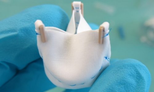
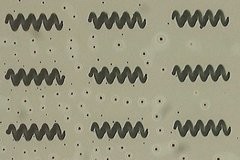
“We’ve printed the world’s smallest stent with features that are 40 times smaller than any produced to date,” says Carmela De Marco, lead author of the study and Marie Skłodowska-Curie fellow in Bradley Nelson’s research group. The group calls the method they’ve developed indirect 4D printing. They use heat from a laser beam to cut a three-dimensional template – a 3D negative – into a micromould layer that can be dissolved with a solvent. Next, they fill the negative with a shape-memory polymer and set the structure using UV light. In the final step, they dissolve the template in a solvent bath and the three-dimensional stent is finished.
Picture: Carmela de Marco / ETH Zurich
It’s the stent’s shape-memory properties that give it its fourth dimension. Even if the material is deformed, it remembers its original shape and returns to this shape when warm. “The shape-memory polymer is suitable for treating urethral strictures. When compressed, the stent can be pushed through the affected area. Then, once in place, it returns to its original shape and widens the constricted area of the urinary tract,” De Bernardis says.
But the stents are still a long way from finding real-world application. Before human studies can be conducted to show whether they are suitable for helping children with congenital urinary tract defects, the stents must first be tested in animal models. However the initial findings are promising, “We firmly believe that our results can open the door to the development of new tools for minimally invasive surgery,” De Marco says.
Source: ETH Zurich
09.08.2019


