State of the art technology for biomedical imaging
A convenient technology to quantify three-dimensional (3D) morphological features would have widespread applications in biomedical research. Now, researchers from Umea University in Sweden presented a new method for 3D imaging and quantification of biological preparations ten times larger than the limit for traditional confocal microscopes.
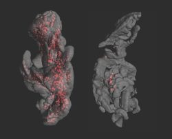
OPT imaged adult pancreata from a wild-type (left) and overt diabetic NOD mouse (right) depicting insulin labeled Islets of Langerhans (red). The specimen are approx. 1.3 x 0.7 cm.
(Umea Center for Molecular Medicine)
(Umea Center for Molecular Medicine)
Traditional biological imaging techniques are limited by several factors, such as the optical properties of the tissue and access to biological markers. In this connection, a major challenge has been the creation of three –dimensional images of the expressions of specific genes and proteins in large biological preparations. It has been equally complicated to try to measure the mass/volume of cells or structures that express a specific protein in a specific organ for example.
A study published in the journal Nature Methods (Tomographic molecular imaging and 3D quantification within adult mouse organs, Jan;(1):31-3, 2007. Epub Dec 3, 2006), now presented a solution based on Optical Projection Tomography (OPT). The study took place under the direction of Associate Professor Ulf Ahlgren from the Umea Center for Molecular Medicine in collaboration with with Dr. James Sharpe from the Centre for Genomic Regulation in Barcelona and Professor Dan Holmbert from the Department of Medical Bioscience, Umea University.
Based on combined improvements in sample preparation, tomographic imaging and computational processing, the researchers present a procedure that makes it possible to create 3D images of specifically dyed preparation that are one centimetre of size, eg. adult mouse organs.
Moreover, the method can be used to automatically measure the number and volume of dyed structures in large biological preparations without needing biological marker substances but only antibodies that are in routine use in many research laboratories.
The potential of this technique was demonstrated by following the degradation of insulin-producing cells in intact pancreases from a mouse model for type-1 diabetes. In this case the team around Ulf Ahlgren showed a direct connection between the volume of the remaining insulin-producing cells and the development of symptoms of diabetes.
For further information, please have a look at http://ucmm.cs.it-norr.com/default.asp?id=1007&PTID=&refid=1018
01.04.2007
More on the subject: