Neuroimaging Technique Identifies Patients at Risk for Alzheimer’s Disease in Healthy Brains
How the brain changes with age is not well-characterized and even less is known about the factors influencing the rate of brain aging. Brain imaging can offer a window into risk assessment into for diseases such as Alzheimer’s disease. A recent study demonstrated that genetic risk is expressed in the brains of even those who are healthy, but carry some risk for AD.

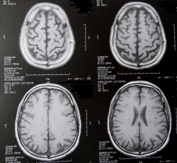
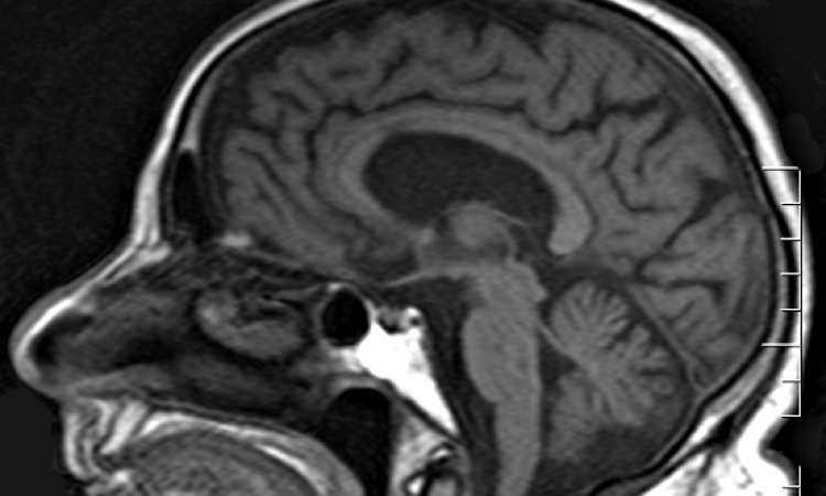
The results of this study were published in the November 2009 issue of the Journal of Alzheimer’s Disease (AD). Investigators used automated neuroimaging analysis with voxel-based magnetic resonance (MRI) imaging diffusion tensor imaging (DTI) techniques to characterize the impact of an AD-risk gene, apolipoprotein E (ApoE4), on gray and white matter in the brains of cognitively healthy elderly from the KU [University of Kansas] Brain Aging Project.
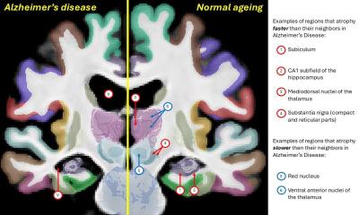
The investigators discovered that healthy elderly individuals carrying a risk-allele of the ApoE4 gene had reduced cognitive performance, decreased brain volume in the hippocampus, and amygdala (regions important for memory processing), and decreased white matter integrity in limbic regions. These types of brain changes are also found in people with AD. Therefore, brain changes, typically found in AD patients, are also evident in nondemented individuals who have a genetic risk of later developing AD.
Lead investigator, Robyn Honea, D.Phil., research assistant professor, University of Kansas School of Medicine (Wichita, USA), department of neurology, Alzheimer’s and Memory Group, commented, “It is important to note that findings of imaging phenotypes of risk variants, such as with this gene, have been shown in a number of studies. The unique element of our study is that we used several new neuroimaging analysis techniques. In addition, the individuals in our study have been well-characterized in a clinical setting.”
This research was conducted in the laboratory of Jeffrey M. Burns, M.D., associate professor in the department of neurology at the University of Kansas Medical Center. He is the director of the Alzheimer and Memory Center and the Alzheimer’s Disease Clinical Research Program. Dr. Burns serves as the lead investigator of the Brain Aging Program.
07.01.2010


