Mystery of complicated plaques
Until recently, physicians believed that stenosis was responsible for strokes. But new investigations show that the composition of complicated plaques are the major cause. Canadian researchers now used 3-D MRI to accuratly detect bleeding by those plaques within the vessel walls.
When major arteries are affected by atherosclerosis, fatty deposits, or plaques, accumulate on the inner lining of the vessel walls. Progression of the disease over time leads to narrowing, restricting blood flow or becoming completely blocked.
Until recently, scientists believed d stenosis, was responsible for most heart attacks or strokes. But new studies have identified the composition of complicated plaques as being a major cause of vascular events and deaths. These complicated plaques are characterized by surface ulcerations, blood clots and bleeding into the vessel wall.
"There's been a major change in our research," said Alan R. Moody of the University of Toronto. "We now know that the composition of carotid artery plaque is likely to be more predictive of future stroke events than the amount of stenosis in the vessel."
In the study, conducted at the Sunnybrook Health Sciences Centre in Toronto, researchers performed 3-D MRI on the carotid arteries of 11 patients, age 69 to 81. Complicated plaques were then surgically removed from the patients' diseased arteries and analyzed under a microscope.
The research team found strong agreement between the lesions identified by MRI as complicated plaques and the microscopic analysis of the tissue samples.
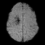
"With high spatial resolution 3-D MRI, we are able to noninvasively analyze the tissue within the artery wall and identify small bleeds within rupture-prone plaques that may put patients at risk for future stroke," Dr. Moody said.
According to Dr. Moody, 3-D MRI is a tool that is ideally suited to screen high-risk patients for complicated carotid plaques and to monitor the effects of interventions designed to slow the progress of the atherosclerotic disease. The technique is easy to perform and interpret and takes only a few minutes when added to an MR angiography study, he said.
This article is adapted from the original press release.
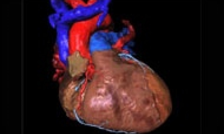
Courtesy of St. Vincent’s Melbourne, Melbourne, Australia
29.09.2008