Molecular MRI
Moving into the clinic?
By Dr Fabian Kiessling, Head of the Division of Molecular Imaging, Department of Biophysics and Medical Radiation Physics, German Cancer Research Centre (DKFZ)
Dr Fabian Kiessling
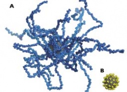
Superparamagnetic iron oxide particles (SPIO) and ultra-small superparamagnetic iron oxide particles (USPIO), amplify the MR signal considerably, compared with established Gd-based contrast agents, due to the crystalline structure of thousands of iron ions. SPIO and USPIO, which accumulate intracellularly, are usually used for specific and cellular MR imaging. These dextran or carboxydextran coated SPIO regularly show high hepatic uptake and are clinically used for liver imaging (e.g. Resovist, Schering, Berlin). For specific imaging it is desirable that the unspecific uptake by the liver and the RES is low and the size of the particles is small. Among those particles are citrate coated, very small, superparamagnetic particles (VSOP), developed by the Ferropharm GmbH (Teltow, Germany; fig. 1). These particles show good biocompatibility and have already entered clinical evaluation as contrast agents for MR-angiography and cardiac MRI.
Nano AG is a multicentre project with partners from the industry (Siemens, Erlangen, Germany; Ferropharm GmbH, Teltow, Germany; Schering AG, Berlin, Germany; Mevis GmbH, Bremen, Germany) and scientific institutions (Charité Universitãtsmedizin Berlin, Germany; University of Freiburg, Germany; German Cancer Research Centre, Heidelberg, Germany) funded by the German Ministry for Education and Research (BMBF) and co-ordinated by Arne Hengerer from Siemens AG (Erlangen, Germany).
The primary aim of Nano AG is to develop target specific VSOP and imaging strategies to visualise dangerous vulnerable plaques in arteriosclerosis using MRI. In addition, it will be investigated whether these contrast agents can also be used translationally, e.g. to detect angiogenesis in cancer. In contrast to most previous projects, it is not the aim of Nano AG to show only feasibility of molecular MR-imaging concepts, but to develop specific contrast agents that can subsequently be applied in patients. Superparamagnetic particles, which are used for this purpose, must fulfil important requirements: First, to be non toxic and highly biocompatible. Second, high T1- and/or T2-relaxivity are required to allow visualisation of the targets in vivo by MRI. Third, small particle sizes are desired to improve extravasation, target binding in vivo and if possible renal elimination.
Nano_AG project particles will be provided by Ferropharm GmbH, which specialises in superparamagnetic iron oxide particles for MRI. In the course of the project the physical characteristics and pharmacokinetic properties of the particles will be tailored to the particular application. VSOP will be functionalised by coupling them to ligands to make them bind to arteriosclerotic plaques. Finally MR-imaging techniques will be optimised and postprocessing algorithms for the MR-data will be developed.
Summary - Nano_AG is a unique interdisciplinary project that combines world-class expertise in probe design, application development and engineering in a cross industry collaboration including renowned academic sites. Specific MR contrast agents will be synthesised for the imaging of early thrombotic plaques. The virtual team will ensure that these functionalised VSOP fulfil the regulations for clinical contrast agents and it is the declared aim that these specific compounds are ready for clinical evaluation after funding ends. Thereby the consortium aims to help in early diagnostics and risk stratification of the ‘vulnerable patient’ by providing a diagnostic solution (contrast agent and imaging hardware & software) for a hitherto unmet medical need.
02.08.2006
More on the subject: