Magnetic resonance mammography
Many diagnostic advantages and more to come
Women in families with a history of breast cancer have a significantly higher risk of developing the disease.

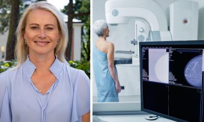
‘For these patients MRM (magnetic resonance mammography), which is an MRI of the breast, is a very effective procedure to detect malignant tissue changes at an early stage,’ said Evelyn Wenkel MD, who is using Siemens’ Magnetom Espree for magnetic resonance mammography (MRM) at the Radiological Institute at Erlangen University. ‘These patients require a particularly close-meshed net of early screening measures. MRI is clearly more unerring here than other diagnostic procedures – and compared to conventional mammography it also works without ionising radiation. ‘To achieve sufficiently adequate image quality in breast diagnostics we need to work with a field strength of at least 1.5 Tesla. Additionally, we need special, bilateral breast coils or, even better, multi-channel coils. With the sequences the examiner needs to ensure that the resolution regarding the recognisability of details is large and that the sequences are fast enough to show the accumulation of the contrast medium in a tumour. A malignant tumour, for instance, would normally absorb the contrast medium quickly, a benign tumour more slowly.’
Dr Wenkel is watching the race for increasingly larger field strengths with reservations – at least in terms of breast cancer diagnostics: ‘You can also improve the quality of MRI images through optimised coil and sequencing technology. At the moment, as far as I am aware, there are no randomised studies that prove the superiority of 3.0 or 7 Tesla machines over the 1.5 Tesla MRI scanners used for the diagnosis of breast cancer.’
Compared to sonography, one essential advantage of MRM is the lesser influence of the examining physician on the results, which makes MRM images more objective and easier to reproduce, and also makes it suitable for monitoring and aftercare, she points out. ‘In the past the differentiation between scar tissue and new tumours was always a big challenge, even for experienced diagnosticians. This hurdle is now easier to overcome with the help of MRM.’
MRM, she believes, will continue to gain in importance. However, for the early detection of breast cancer it will initially only be used for high risk patients due to the higher costs compared to traditional mammography and the number of available scanners.
A further application for MRM is in CUP (cancer of unknown primary) syndrome, which experts refer to when secondary tumours are found without the location of the primary tumour being detected. For this difficult diagnostic problem radiologists are pinning a lot of hope on MRM. For Dr Wenkel and her colleagues all the advantages of the procedure have not yet been exhausted. ‘Apart from purely morphological information about the mammary gland and its changes, MRM can deliver information about the biological characteristics of a tumour, such as cell density or biochemical characteristics. Finally, it has been shown that MRM can also significantly impact on surgery planning. For example, if a further tumour is diagnosed in the breast with the help of MRM this is of crucial importance for the operation. In certain cases an MRI can be important before breast surgery, so that all primary tumours can be detected and then removed during one intervention.’
01.05.2009


