Light therapy supports diagnostic imaging
One of Europe's most modern radiology and diagnostics centres opened in Vienna-Donaustadt, Austria, this January.
Built up from around six million euros raised by private investors, and now offering state-of-the-art technology plus medical expertise and patient-oriented services, the centre will readily compete with any university hospital. Christian Pruszinsky, our correspondent in Austria, asked Dr Gunther Alth, the centre’s manager, and Dr Friedrich Vorbeck, how this centre differs from the rest
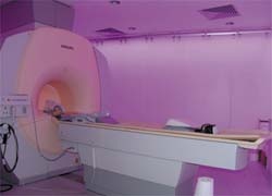
‘In our private centre we try to live a new dimension in diagnostics,’ Dr Alth explained. ‘This means we provide a comprehensive range of equipment of the latest generation for digital X-ray, digital mammography, ultrasound, CT, MRI and radiotherapy. Moreover, we are the first in Europe to offer light therapy support – which our patients embrace enthusiastically.’
Asked how that novel system works, he pointed out that many patients feel very uncomfortable during MRI and CT exams, and sometimes they have specific problems - such as claustrophobia. ‘So we opted for Ambient Lighting, a dynamic lighting concept by Philips. It’s designed to create a soothing atmosphere in the examination environment. Light creates wellness is the motto of this concept. A patient being moved through the scanner can look at a harmonious play of pastel colours that soothes and relaxes him, or her, and detracts from the actual examination. The felt scanning time is shorter, the play of colours reduces fear and makes patients feel secure and at ease.’
Asked how the lighting is integrated into a clinical procedure, and whether it resembled a pre-programmed light show in a discotheque, Dr Alth said the interaction of the homogeneous general lighting system and the additional wall-mounted LEDs is controlled manually, to be able to adapt it to individual situations. ‘Another advantage is that we can communicate with patients with hearing problems via colour codes. In MRI or CT scans you often have commands such as “hold your breath” or “resume breathing”. Now, for example, a green light, can tell the patient to breath again, while a red light tells him to hold a breath. That makes life easier not just for patients but also the staff.’
At the clinic, wellness seems to be a leitmotiv – from the reception through the waiting area to the hallways: generous light, pictures, and friendly colours… It’s almost like in an art gallery, I remarked. Dr Alth was pleased. ‘I’m happy that this is your spontaneous impression, because that is exactly what we wanted to achieve with our focus on superior service and where we consciously want to offer an alternative to the often quite depressing hospital routine. And indeed, we opened with a large Prachensky vernissage. Others will follow.’
On 700 m2 the management have installed a state-of-the-art equipment park for imaging diagnostics and plan to handle up to 500 patients daily. ‘That’s quite a task for the medical staff,’ I suggested, but Dr Alth was not fazed. ‘We have a team of top specialists and a board of experts that will support us with complex cases,’ he said reassuringly. ‘So, fast and accurate diagnoses are guaranteed.’
08.03.2007