fMRI may detect brain activity of patients in vegetative state
British researchers detected near-normal brain activity in patients who are believed to be in a persistent vegetative state (PVS) by using functional MRI. The findings may help doctors' to diagnose patients that might have a chance for recovery, but the researchers warn to over-intertpret their resulsts.

Severe brain damage may result in a condition of persistent vegetative state (PVS): patients breath and open their eyes, they have reflexes but there is no indication for consciousness. The care concept of basic stimulation shows some effect but usually a more than 2 years lasting PVS has virtually no hope of recovery.
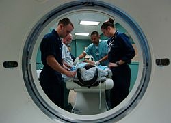
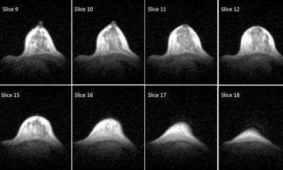
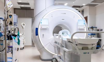
Dr Adrian Owen, Senior Scientist and Assistant Director at Britain’s Cambridge University, and colleagues used funtional MRI (fMRI) to visualize the real-time brain activity of patients in persistent vegetative state. “We observed twelve patients and asked them to imagine playing tennis,” reports Owen in the Archives of Neurology. “Two scans showed brain activity nearly identical to that of healthy people.”
“The first patient is improving. About six months after we scanned her, she started to show the earliest signs of improvement. She is now in a minimally conscious state,” explains Owen. The team does not want to overvalue the findings although it may be possible to prognose chances of recovery. It appears that PVS caused by brain injury after trauma is more likely to recover than PVS caused by lack of oxygen.
“But we don’t want to raise false hopes or make people think all minimally conscious patients are aware. There is as yet no treatment or intervention that has been empirically tested and shown to be beneficial in this patient group,” concludes Dr Owen.
17.08.2007


