Elastography supports diagnosis and clinical monitoring
With the possibilities of tissue elasticity measuring the diagnostic use of ultrasound has recently increased enormously; the opportunities and limits of this technology and its areas of application are continuously growing.
Dr Dirk André Clevert of the Institute for Clinical Radiology at Ludwig Maximilian University, Munich, explains what this means for daily clinical routine.
In 2004, the ultrasound activities at the Grosshardern Surgical Clinic and Polyclinic at the Institute for Clinical Radiology, and the Grosshardern II Medical Clinic and Polyclinic were merged and the Interdisciplinary Ultrasound Centre was founded at the University Hospital in Munich-Grosshardern.
Since then, there has been a significant increase in the number of indications and a broader range of applications for ultrasound. Overall, the partner clinics carry out about 18,000 diagnostic examinations annually at the centre, assuring the accumulation of a lot of experience, particularly in imaging diagnostics.
Strain Imaging is a procedure which, for the first time, can assess the stiffness of the tissue alongside the delivery of pure image information. With elastography one generally distinguishes between two procedures – the manual compression of tissue with the transducer to assess superficial lesions, and acoustic radiation force impulse (ARFI) imaging. An acoustically induced pushing pulse produces tissue displacement on a micrometer level. Detection pulses register the displacement along the axis within the ROI. The relative displacement is displayed as an elastogram with gray-scale curves.
This allows the doctor to assess the mechanical characteristics of the deep tissue and recognise changes in the tissue. This automatic elastography procedure increasingly works independent of the examiner, Dr Clevert emphasised. Another big advantage for results interpretation is that a numerical value is obtained that reflects the propagation velocity of the shear waves. This value also enhances the objectivity of the diagnosis.
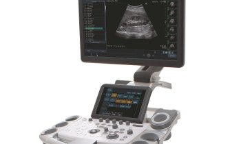
The Siemens system used at the interdisciplinary ultrasound centre has a quality factor that continuously provides the examining doctor with reports about the signal strength in the tissue and therefore with information about elastogram quality. The increasing specificity and sensitivity of elastography has also increased the number of potential indications for this examination. ‘We have been gathering experience with this new ultrasound technology for about a year now,’ said Dr Clevert. ‘Initially, our focus was on liver diagnostics to determine fibrotic and cirrhotic liver changes. Meanwhile, elastography is also now used for other diagnostic indications because of the interdisciplinary character of our centre. It helps us that our system can compare pathologies in the existing B-image with the elastography image due to the dual imaging, and that the automatic B- image recognition (eSie Calcs) can capture these pathological changes and precisely analyse the spread, as well as localisation, in both images.
A new multi-centre study
By the time the elastography system was launched at ECR 2007, Siemens had already published data from the Barr study, which proved that the new ultrasound procedure is particularly suitable to differentiate between normal and malignant breast tissue. At the time, Richard Barr, Professor of Radiology in Ohio/USA, who headed the study, expressed hope that elastography will negate the need for numerous biopsies. Now, Dr Clevert and his colleagues aim to collect further data, along with other universities in a large multi-centre study by examining liver disease with the ARFI method. Initially, to check the reference data, biopsy will be used as the gold standard of tissue analysis.
‘As elastography is usually a complementary procedure to other diagnostic ultrasound procedures, it is important for us to know where exactly a suspected tissue change is located. Our system can control the elastography so precisely, based on an existing B-image, that we can examine the characteristics of the tissue at exactly that point,’ he explained.
Elastography may have the status of a complementary examination procedure that is used when other ultrasound diagnostic procedures have not delivered sufficient, conclusive results, he pointed out, but, independent of the diagnostic applications, this procedure will be able to support therapy and clinical monitoring in the future. ‘A good example is hepatitis; if, six months after the onset of the disease, no normalisation of the transaminases has occurred and HBeAg continues to be detectable, then this disease can be treated with very expensive interferon therapy. However, we know that by far not all patients respond to the administration of interferon. With elastography we can monitor the consistency of the liver tissue and if necessary adjust the therapy scheme at an early stage.’
20.05.2009


