Defying physics
MRI-PET could arrive in 3-4 years
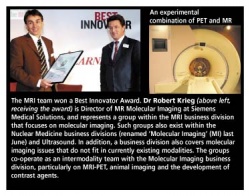
Dr Robert Krieg, Director of MR Molecular Imaging at Siemens Medical Solutions, described, in an interview with Daniela Zimmermann of European Hospital, the limitations of physics and the potential clinical benefits of hybrid technology - and a hitherto hush-hush MRI-PET project

So far, it has been impossible to combine PET and MRI because current PET systems are based on photo multipliers that are incompatible with the magnet. ‘Our approach is to replace the photo multiplier with semiconductor detectors - a bit like a CCD chip in a digital camera,’ Dr Krieg explained. We want to use a semiconductor detector that is magnet-compatible. This new MRI-PET will only work if we really succeed in applying semiconductor technology to PET. So, in that sense, it’s a re-invention of PET. It means we are facing two technological challenges. So far, we have been able to establish the proof of principle - we can prove it works.’
What is a proof of principle?
‘An experiment that proves that a certain method works in principle; based on that proof, a company usually decides whether to proceed with a certain idea and turn it into a real project, or ditch it. Once the proof of principle is established the actual work starts: How can we turn the idea into a functioning system that meets all requirements? This is no longer lab talk, it’s the real world. Realistically, MRI-PET could take three to four years to develop. First we need to tackle several problems: Semiconductors are tailor-made in only a small batch. The production process for these is quite different from that of regular photodiodes. Currently there is only one company, worldwide, that comes close to mastering that process: Hamamatsu. And we have no idea yet how stable the process is.
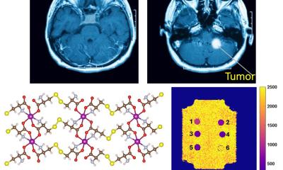
‘Then there is the issue of temperature stability. We put real stress on our gradient coil. If the temperature fluctuations resulting from that high-performance use influence the PET detector the image quality will suffer. We have to solve that problem. In addition to technical questions, we also try to look at this entire issue from a holistic viewpoint. In other words we also ask what clinical benefits the MRI-PET will have and we collect cases that show findings that a PET-CT would not detect. For example, we detected a breast carcinoma in a 73-year old woman, which a CT could not have detected with such clarity. One objective of our clinical co-operation is finding cases that prove that indeed there is an added clinical value and that there are patient groups who would profit from MRI-PET. We must also ask ourselves whether this additional market not only exists but whether it is big enough to warrant the expense.’
Those are the first steps, and then...?
‘Then we will reach the really innovative part. We still have a long way to go and many colleagues will put their shoulder to the wheel. We will celebrate progress and suffer setbacks until we finally have a product.
Now that the veil has been lifted on this project, aren’t you afraid that competitors will jump on the bandwagon?
‘We have a head start in terms of knowledge. It will be difficult for the others to catch up. Look at the synergies: Typically engineers who deal with PET haven’t a clue about MRI. In this we are quite a bit ahead, because we have all the experts sitting around one table: colleagues who have worked on semiconductor detectors for years, as well as excellent MRI physicists. Getting these experts together - that’s the real work of art. The results so far are definitely exhilarating and this motivates the team - and enthusiasm creates wings. Once you have wings, you fly.’
07.08.2006


