

Source: Shutterstock/Iulia Ghimisli
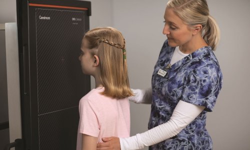
Sponsored • Intensive care
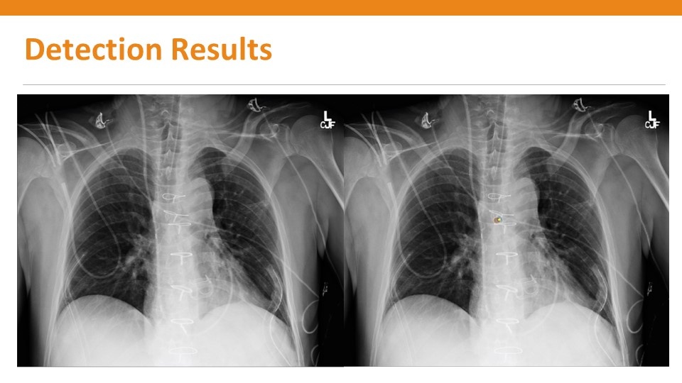
Deep learning software helps to locate the carina
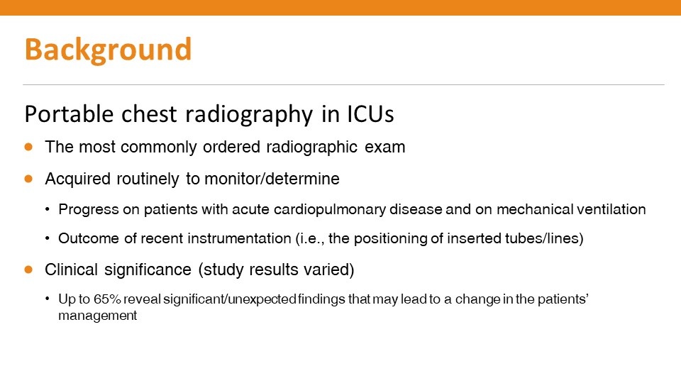
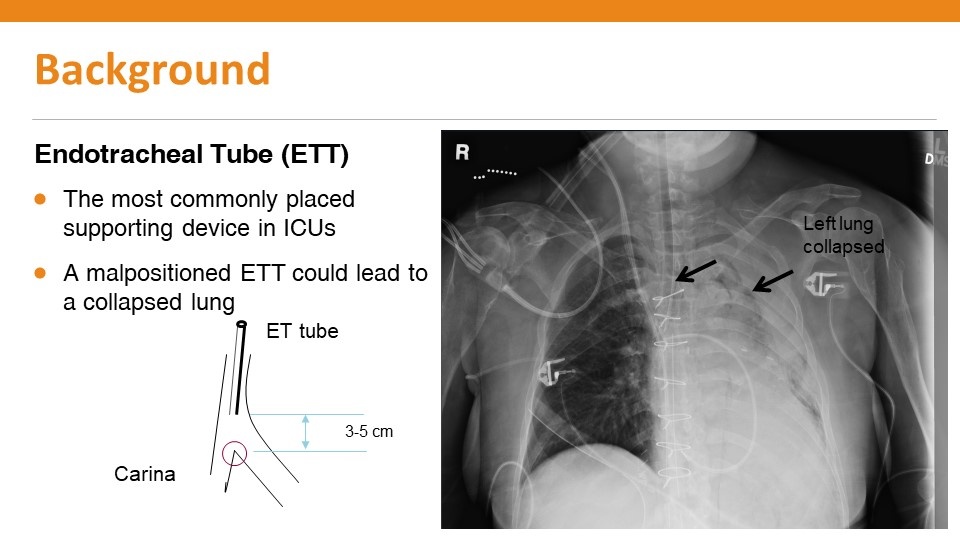
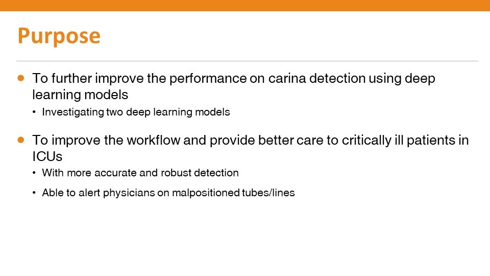
The ability to accurately check the position of the endotracheal tube for patients in intensive care units is crucial to their wellbeing and safe treatment. A pivotal element of this lies in identifying the position of the carina, a ridge of cartilage in the trachea that occurs between the division of the two main bronchi.
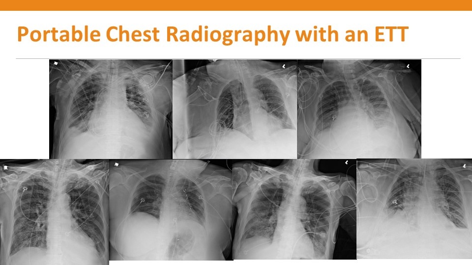
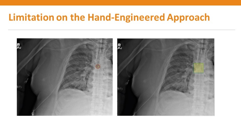
Yet highlighting its location often proves problematic on portable chest x-rays because of low image contrast and ‘noise’ in the region of observation and requires high levels of expertise and experience. Any error can have a negative impact on the patient’s welfare. To support radiologists and intensive care physicians in this, Carestream Health has devised software that it believes can pinpoint more accurately the precise position of the carina and assist in the positioning of the endotracheal (ET) tube.
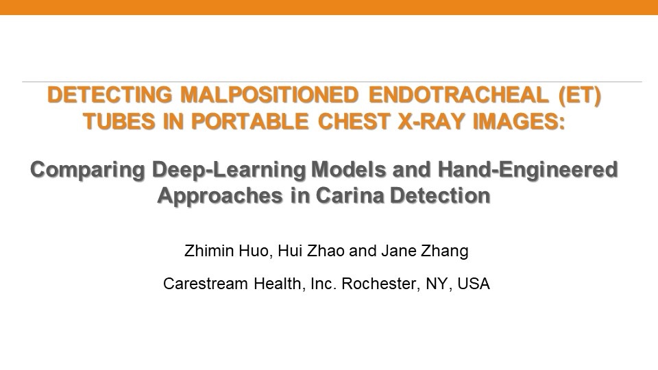
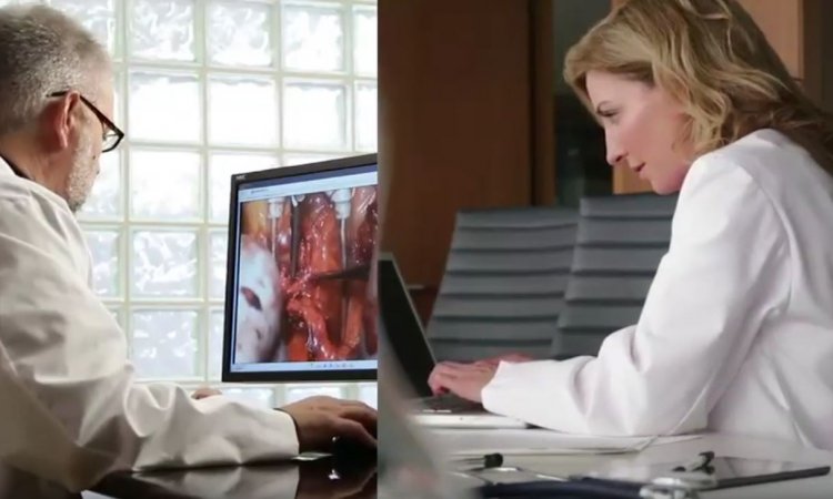
The benefits of the software are among medical imaging advances the company will outline at the Radiological Society of North America (RSNA) annual conference in a session on November 27 entitled “Detecting Mal-Positioned Endotracheal Tubes in Portable Chest X-ray Images: Comparing Deep Learning Models with Hand-Engineered Approach on Carina Detection.” Ahead of the congress, Jim Sehnert, director of research and innovations, Carestream Health, discussed some of the main points. He explained that it can often be difficult to find the carina on an x-ray image and subsequently to place the endotracheal tube, particularly in the setting of the Intensive Care Unit (ICU) where most images are acquired without the use of an anti-scanner grid.
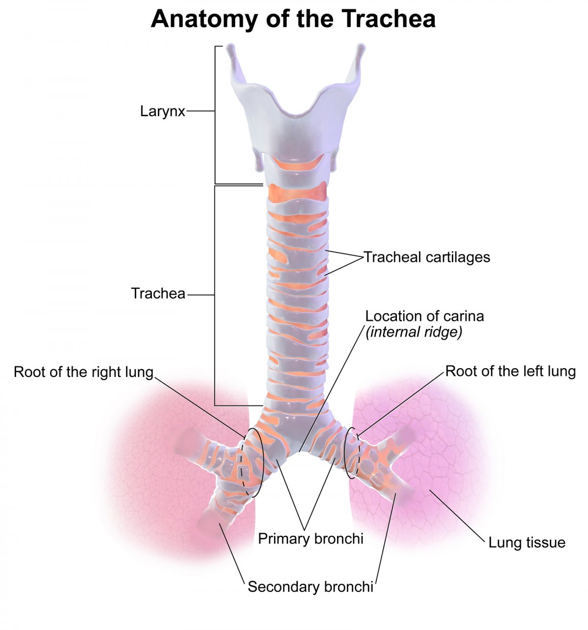
Blausen.com staff (2014). "Medical gallery of Blausen Medical 2014". WikiJournal of Medicine 1 (2). DOI:10.15347/wjm/2014.010. ISSN 2002-4436., Blausen 0865 TracheaAnatomy, CC BY 3.0
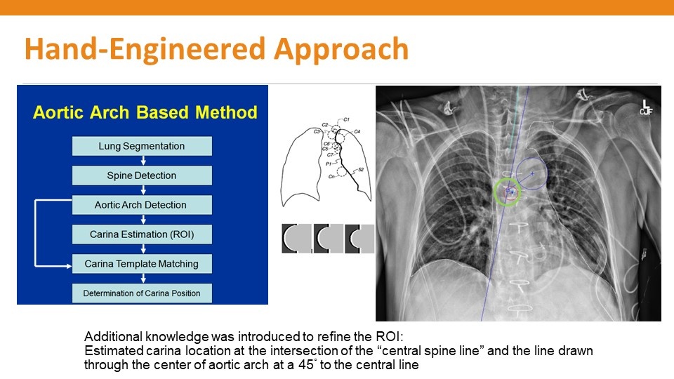
Traditionally, the carina has been located by the radiologist either by taking the position as the middle of the T4-T5 interspace; or by using the Dee Method, which involves identifying the aortic arch and then drawing a line inferomedially through the middle of the arch at a 45-degree angle to the midline. The intersection of the midline and the diagonal line is the likely position of the carina.
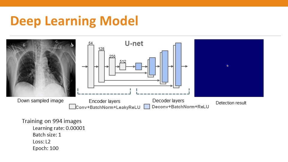
From an imaging science perspective, Sehnert said an algorithm can be created through “heuristic processing” based on the method of Dee and then use additional processing designed to identify the carina by its shape to fine tune the final positional estimate. Yet with deep learning, the process moves away from heuristics. “These complex medical models tend to be more robust against failure than a heuristic example composed of many steps and developed on a limited number of examples,” said Sehnert. “Deep learning learns everything internally, requires no intuition whatsoever, and just requires a lot of data that has ‘truth’ associated with it.”
In addition to ‘truthing’ by the radiologists, in the more difficult cases image scientists can use the specific tools at their disposal – such as enlarging the image, adjusting the contrasts, or applying sharpening filters - to better visualize the location to establish truth. He added: “Some of those tools are available on a PACS viewing station but can go unused because of the workflow issues associated with the radiologists.”
Carestream Health is advocating the benefits of the deep learning approach through the new solution it believes will deliver improved accuracy. Sehnert explained: “Deep learning is applied to the image as it is acquired and the information can be potentially put into a structured report and then delivered to the PACS workstation and displayed as an overlay onto the image, which the radiologist can use as a reference point to gauge the location of the carina.” That can then be used to assess the position of the ET tube relative to the carina, facilitating early detection and prompt correction of improper position of the endotracheal tube as necessary.
The software also detects the location of the ET tube, and the carina, and can report the measurement automatically.
Whether it is the radiologist, or intensive care specialist, reading the report will depend on where the software solution is located. Sehnert said it is possible to load the software into the portable imaging device in the ICU. “In this case,” he continued, “the algorithm can inform the technologist on the spot whether or not the tube is mal-positioned and then inform the intensivist that an action needs to be performed to properly position the ET tube.”
It is also possible to perform the process with the software in another location with the image pushed from the portable imaging device into the PACS system for a radiologist to read remotely. “However, we would prefer the software to work on the portable acquisition device because it gives an answer at the point of care and mean problems can be addressed as they arise. Waiting for the radiologist to give that reading could be some hours.”
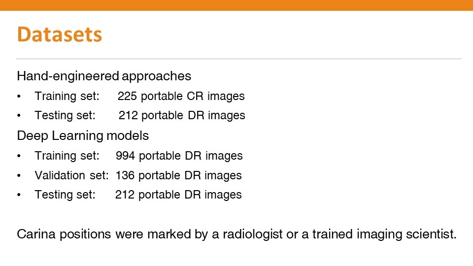
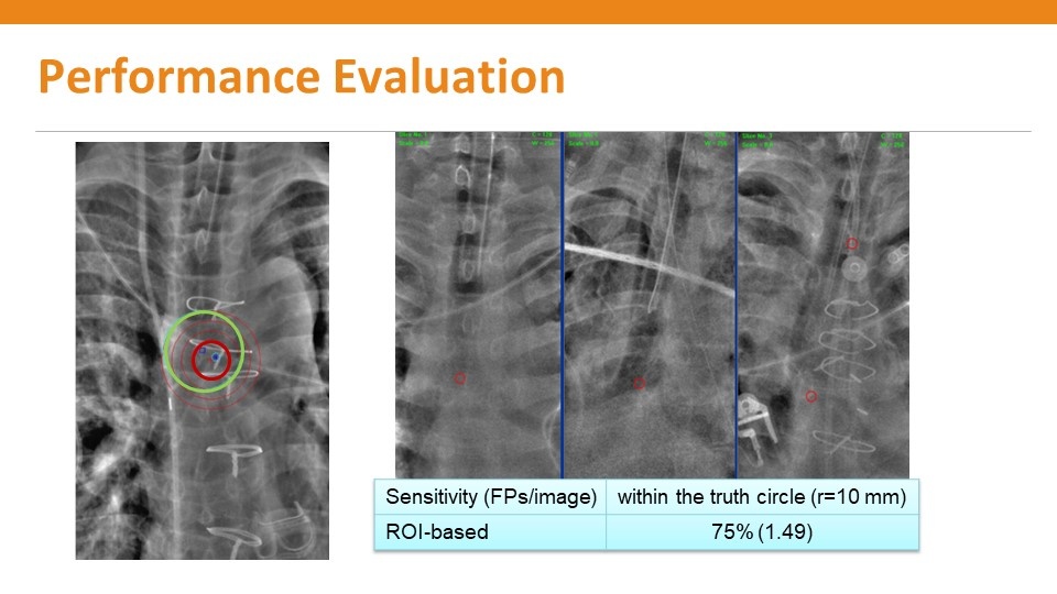
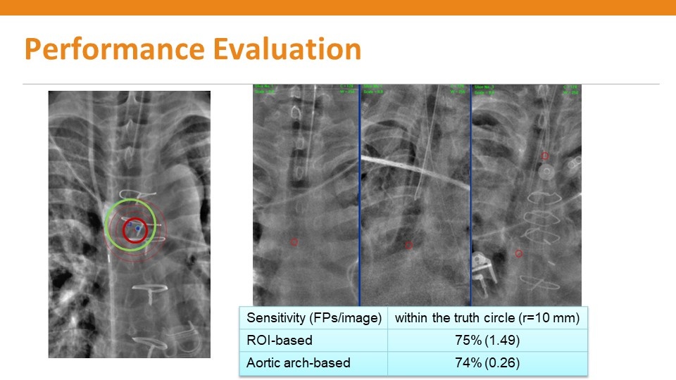
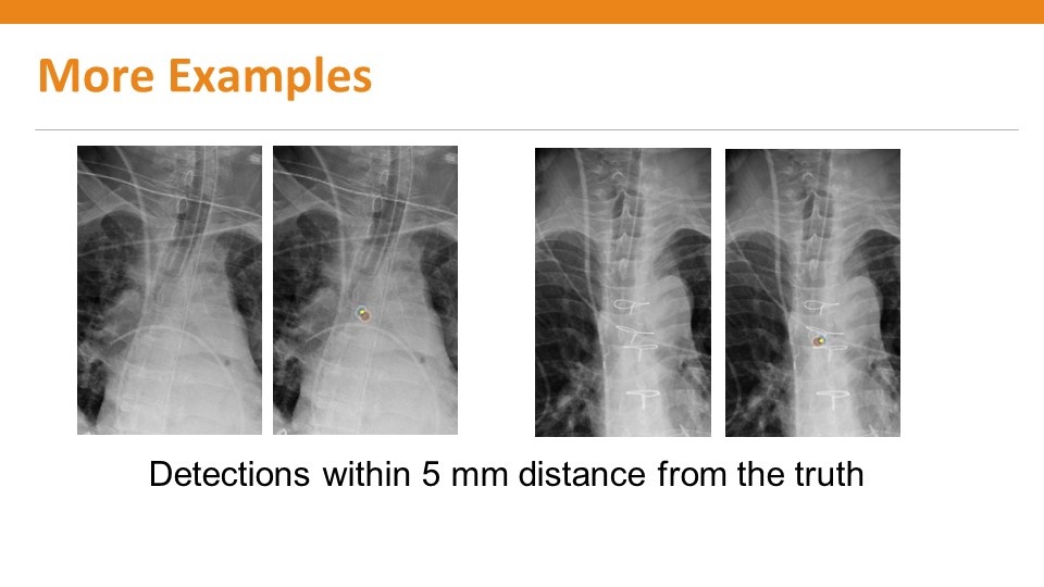
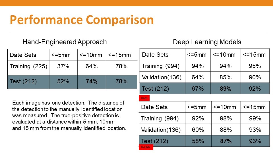
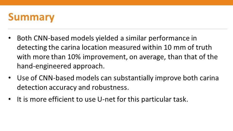
The software has been developed by creating an algorithm from tens of thousands of chest radiographs. In assessing the performance of the algorithm, Sehnert said the development team consider an estimate of within 1cm from the true position of the carina to be accurate. Using this metric on 212 test cases, they found the manual algorithm identified the position of the carina in about 74% of cases whereas for the deep learning approach it was 89%.
Session: Detecting Mal-Positioned Endotracheal Tubes in Portable Chest X-ray Images: Comparing Deep Learning Models with Hand-Engineered Approach on Carina Detection, RSNA, Tuesday, November 27, 11:10-11:20am. Authors are: Z. Huo, J. Zhang and H. Zhao.
25.11.2018