Imaging equipment
CT and MRI-compatible compression harness
The DynaWell L-Spine, a compression harness that axially loads the lumbar spine in the supine position, has no magnetic parts, so can be used with most CT and MRI scanners.
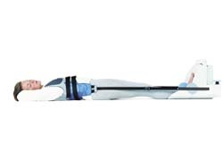
Closely resembling a spinal column when loaded with a patient in an upright position, this compact, lightweight system includes a compression vest attached by straps to a foot-driven compression device. Pressure exerted by the system is not placed directly on a patient’s shoulders, says DynaWell International.
When examining patients in an unloaded, psoas-relaxed position (PRP), narrowing of the spinal canal could remain undetected. However, when examined in a slightly extended position, during axial compression (ACE), pathologic features can become more visible. Use of the spinal-compression device helps detect encroachment of the spinal canal, associated with pathological changes (e.g. stenosis, disc herniation, intraspinal synovial cysts).
Clinical studies have now shown that a more specific and valid diagnosis can be achieved by using a lumbar-spine compression system rather than a traditional non-loaded PRP. Published research results indicate that, by using the compression system, the likelihood of spotting a stenotic situation in the spinal canal increased by 60-70%, compared with detection during the usual unloaded examination. Further clinical studies are taking place at the University of Aberdeen (Scotland), Mannheim (Germany) and Rochester (USA).
[Update] The manufacturer of the harness, DynaWell Diagnostics, seems to have ceased operation.
30.04.2003