An affair of the heart
ESCR 2009
The heart of cardiac radiology set the rhythm for this October's 10th Annual Meeting of the European Society of Cardiac Radiology (ESCR) in Leipzig, when state-of-the art technology and progress in cardiac imaging were introduced alongside an educational programme that catered for experienced as well as novice cardiac radiologists
Explaining the aim of ESCR 2009, its president, Professor Michael Rees MD (School of Medical Sciences, Bangor University, Wales) said: ‘As the technology for cardiac imaging has rapidly developed so has the need to train and educate radiologists who have an expertise in the clinical and scientific aspects of cardiac imaging. This is one of the prime missions of our society.
‘As there is a steady growth of other professionals making a significant contribution to cardiac radiology, we extended our congress programme with special sections catering for the interests of radiographers and technologists this year. Moreover, the ESCR is now actively seeking to develop and involve national cardiac radiology societies. A forum for these societies is under development.
‘The success of cardiac radiology in the future will depend not only on a strong and vibrant European Society but the development and interaction between our society and the growing number of national societies that have an involvement in cardiac radiology.’
Prof. Rees said that national efforts in cardiac radiology have been considerable. Different countries are indeed at different stages with their developments in programmes, training and research, but EU countries like Greece, Portugal or Spain are catching up rapidly with the pioneers, such as Germany, The Netherlands and Italy. The focus on international collaboration during ESCR 2009 went even a little further with South Korea as a special guest. ‘Our Asian-Pacific colleagues have a lot of experience with new CT and MRI machines because a lot of medical engineering comes from this region,’ he explained.
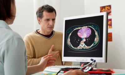
Therefore, educational sessions and a hands-on workshop around Cardiac CT and MRI were constituted as a priority of the meeting. But, when does the cardiac radiologist use what? ‘At the moment, cardiac CT is mainly used in two areas. One is CT scanning for calcium score to check for the build-up of calcium in plaque on the walls of the coronary arteries,’ he said. ‘The other one is to look at coronary artery vessels and bypass vessels after injection of contrast agents to evaluate the coronary artery vessels directly for stenosis or closure.’
Due to the recent advances in CT scanners – faster machines, faster examinations – the range of conditions under which patients can be examined is broadening, e.g. for acute patients in the emergency setting who cannot hold their breath. ‘Cardiac MR is on the other hand the gold standard for the volumetric and functional analysis of the heart and allows for a detailed tissue characterisation of the heart muscle (myocardium). It is especially useful for the detection of scaring and viability after a myocardial infarction.’
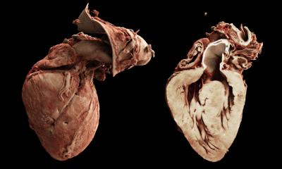
In his own lecture, Prof. Rees focused sharply on Electron Beam Computerised Tomography (EBCT) as a catalyst for cardiac imaging, Coronary artery calcification EBCT scanning is a test that affords the opportunity to determine very accurately whether or not coronary artery disease is present in asymptomatic individuals. It uses an electron gun and stationary tungsten 'target' rather than a standard X-ray tube to generate X-rays, permitting very rapid scanning times.
EBCT, an invention from the ’80s, was the subject of a keynote lecture by its inventor, Douglas Boyd who spoke of the past as well as future developments. Outlining the new he explained that, because these machines can be made much smaller, they could gain higher significance in competition with conventional multislice CT in the future
12.11.2009