Advances in medical imaging: Moving on – from measuring to planning
Workshop: 12-16 March
University of Technology (RWTH) Aachen, Germany
Prior to the imaging workshop , Meike Lerner asked programme organiser, Professor Thomas M Deserno (Dept. of Medical Informatics, RWTH) about current developments today and how they affect future input from imaging manufacturers.
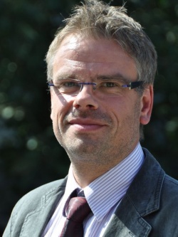
‘So far, the objective of medical image processing has been to generate reproducible and quantifiable information from images and utilise it purely for diagnostic purposes. Application-specific aspects were not that important,’ he pointed out, adding that this is now changing. ‘Medical imaging permeates patient care, treatment planning and therapy itself. Technologically, this means that, whilst meeting the standard requirements, new algorithms primarily have to deliver real-time information – such as directly to the monitor in the operating theatre.’ Overall, he explained, there are four areas for future image processing: Multidimensional image formation, computer- assisted diagnosis (CAD), intervention planning and treatment (computer-assisted surgery). ‘We’re already on the right track with the first two, as shown in computeraided diagnosis in mammography, where doctor and computer work really well together now. The challenge for the future will be to extend this methodology to other applications.
This requires the evaluation and verification of large data sets in a large variety of different areas to develop the respective algorithms.’ Does the oft-quoted ‘data tsunami’ determine future development? ‘No, it is less a case of the amount of data to be processed – storage is not such an exclusive commodity any more; it’s more to do with the technical implementation. On its own, data is of little use if it is not linked to a validated foundation. We can already filter out reference images from other patients, based on local image patterns, and include these in the diagnosis. However, the question is, to what extent the medical information on which these are based, i.e. a doctor’s letter and diagnosis reading, are really valid and if they can be automatically evaluated via a computer.’
Intervention planning
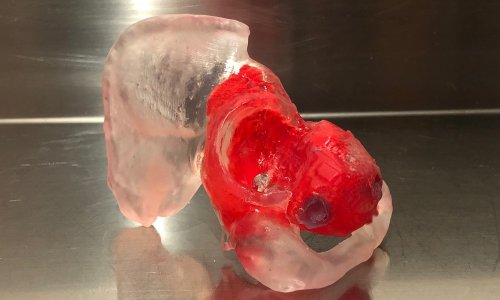
Using a liver transplant as an example, Prof. Deserno said that, for successful treatment in the case of a split – a live organ donation, for example – it is essential that both parts remain functional. ‘The arteries, portal vein and bile ducts can only be dissected at the periphery. For the best possible surgical preparation all liver segments and these trunks have to segmented and visualised in 3-D, so that, on the one hand, certain partial volumes can be calculated precisely via computer and, on
the other, the surgeon is shown the patient’s morphology. This requirement goes beyond pure diagnostics, because the focus is not so much on what the local vessels are like. To put it more abstractly, we leave the level of data and move on to the level of modelling, which shows the surgeon the
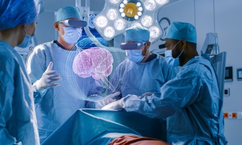
essential facts. This approach presents very new challenges for the algorithms.’ Here, he added, is where the fourth point comes in – CAD -- because the modelling information must be directly and adequately available in the operating theatre. ‘Here we are dealing with questions such as how the data material is represented, or how the methods of augmented or virtual reality can be utilised to give the surgeon additional information during the procedure.’ How far off are such developments? ‘The transitionsare smooth -- as CAD shows, for example. For a few years there’s already been intensive research in all areas – though not really with a lot of steam. Therefore, an important step towards the future is that the necessity of this research is now being acknowledged, and that these topics are placed at the top of the agenda. Our workshop not only reflects this trend but actively promotes it.’ Ulla Schmidt, former German Minister of Health, will open the 12th workshop on medical image processing, for which speakers include Professor Bernd Jähne (Heidelberg) and Professor Axel Wismüller (Rochester, NY).
Details: www.bvm-workshop.org
04.03.2010