A new imaging system for cancer
Accurate diagnostic analysis and staging of cancer of the bile duct still remains a challenge. According to a new study from Germany a new imaging system called Cellvizio allows physicians to examine tissue at the cellular level from inside the body may now enable them to diagnose one of the most difficult cancers to detect.
Cholangiocarcinoma is a cancer of the bile ducts, which drain bile from the liver into the small intestine. With an annual incidence rate of one to two cases per 100,000 in the Western world, this disease has been steadily increasing over the past several decades. Risk factors include inflammation of the bile ducts and liver malfunctions. Symptoms include jaundice, weight loss and generalized itching. This disease is diagnosed through a combination of blood tests, imaging, endoscopy and sometimes surgical exploration. To date, surgery is the only potentially curative treatment.
But according to a new study in the September 2008 issue of the journal Clinical Gastroenterology and Hepatology, a new imaging system that allows physicians to examine tissue at the cellular level from inside the body might help them to a more effectively diagnose .
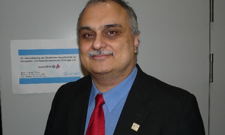
"Accurate diagnosis and staging of cancer of the bile duct remains a challenge despite advanced imaging methods and the introduction of tissue sampling methods," said lead investigator Alexander Meining of the Technical University of Munich. "Adding this non-invasive tool that allows us to view abnormal bile duct tissue while it's still inside the body increases diagnostic accuracy of cancer of the bile duct."
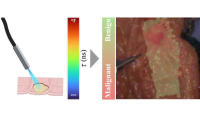
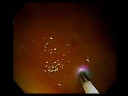
Dr. Meining and his colleagues conducted the 14-patient study to evaluate the ability of the Cellvizio confocal microscopy system to detect neoplasia (an abnormal growth of cells that can be benign, pre-cancer or cancer) in biliary tract tissue by examining tissue at the cellular level during Endoscopic Retrograde Cholangiopancreatography (ERCP), a procedure used to diagnose cancer of the bile ducts and pancreas.
They found that Cellvizio predicted neoplasia with a sensitivity of 83%, considerably better than the 50% seen with standard histopathological analysis of biopsied tissue taken from strictures. In addition, Cellvizio predicted neoplasia with an accuracy rate of 86%, which was superior to the 79% accuracy rate of standard histopathology. "We believe Cellvizio also might facilitate the detection of other cancers arising from ductal structures in the pancreas, breast and urogenital area," Dr. Meining added.
The researchers also reported that Cellvizio images captured with the dye fluorescein enabled them to better differentiate between patients with bile duct cancer and those without it by allowing them to see abnormal blood vessels present only in patients with cancer.
The specially designed Cellvizio CholangioFlex Miniprobe enables the Cellvizio System to be used during ERCP procedures and debuted earlier this year at Digestive Disease Week 2008 in May.
Cellvizio is the first and only confocal microscopy system that is compatible with most endoscopes including cholangioscopes and advanced endoscopic modalities. Cellvizio has 510(k) clearance from the Food & Drug Administration and the European CE-Mark for use in the gastrointestinal and pulmonary tracts. Over 1,500 Cellvizio procedures have been completed to date.
11.09.2008