7-TESLA
Mastering the “uncontrollable beast”
By Brenda Marsh, Editor, European Hospital.
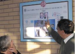
The Sir Peter Mansfield Magnetic Resonance Centre crowns a slope at the top of the University of Nottingham campus – a fitting position, for it was here, over 30 years ago, that Sir Peter Mansfield, Emeritus Professor of Physics, pioneered his work on MRI, which led to the development of the first MRI scanner, revolutionised diagnoses, and resulted in a Nobel Prize, shared with Paul C Lauterbur (Univ. of Illinois, USA) in 2003.
Today, the centre is still the vanguard of MRI research. It was also the first to install the then most powerful magnet - the 7-Tesla - that is so strong, it needs to be contained within 230 tonnes of iron cladding, to protect people from its field.
Might it present any physical dangers, I asked Professor Peter Morris, head of the research centre. ‘There is no effect - that’s my message. If people move through the magnetic field quickly, some may lose balance, or feel nauseous, particularly if moving from 2-T to 5-T across the face, so they’re moved into the scanner very slowly. Once in the middle, the field is uniform and they’re fine! Each time the field goes up the same story emerges, but people don’t worry about it.’ Currently, the platform is manually driven; in the future it will be motor-driven - slowly.
Could 7-Ts enter our hospitals? ‘Far from being the uncontrollable beast, the 7-T works as it’s delivered – producing really great images, and different ones from those obtained at lower Ts, so what it’s giving is new. At first we expected to do as much as five year’s work to get decent images but, from day one, our scientists were surprised at the images.’ However, there is much to be done - and hurdles to jump.
Of the centre’s c. 50 employees, 17 work on Tesla research, under Prof. Morris’s guidance. ‘We are not on our own,’ he says. ‘It’s a very good team.’ About eight Philips researchers - some in Cleveland, Ohio, where the 7-Ts are built - give back-up. During teleconferences once or twice monthly, they work through any problems together, and assess achievements, decide next steps, and discuss timelines for more installations.
As for cost, when quoting someone’s formula: the system’s around a million dollars per tesla! Prof. Morris smiles. That’s not just for the magnet, he reminds me. This beast needs big housing.
In terms of research, although this is a full-body system, and the team’s keener desires might lie in capturing functional activities, as for most research centres with a 7-T, work focuses on static areas such as the brain. Currently they are developing a head-only coil because, to get a better image, it’s more efficient to match the coil to the size of the region being examined.
For multiple sclerosis, there is rather poor correlation between the extent of the disease lesions seen in scans and the clinical symptoms. ‘We know that quite a lot of these lesions have been missed, and hope the sort of contrast we can get at certain fields may actually reveal these,’ Prof. Morris explains. The 7-T brain images have a resolution of .6 of a millimetre, so each image element is .6 x .6 x .6 deep, providing very high resolution. ‘MS patients typically have lesions in the white matter, but there are probably some in the grey areas surrounding it, and want enough resolution to see what’s happening in the outer cortex. Every region of the brain is covered, in .6mm tubes, so we can pick out any of it we want. We are very very pleased with the kinds of contrast we can get - and we get a little bit of functional work.’
Viewing the centre’s images is like exploring deep, mysterious valleys on another planet. A scan, on the sagittal plane, reveals the base of the brain and visual cortex. ‘You can actually see some of the fibres in the little fibre bundles leading from the functional areas, and that’s just on a regular sequence,’ he says. ‘The higher field is being perturbed by those fibres in such a way that it makes them visible. You can’t see these at lower fields.’
Of another image he says: ‘You see grey matter structures that normally you cannot tell apart, but in this lower brain area are different shades of grey; we think it’s because you get selective stimulation of iron in those structures. So iron is going to change electrodes and properties and will do it more the higher the field strength. Of course you don’t know it’s iron, but post mortem studies that indicate these structures accumulate it – in some diseases more so.’
Although none of the images are enhanced, in another scan red nuclei are seen as clear dots, dark due to iron content. They are where damage occurs in Parkinson’s disease – one of the neuro-degenerative diseases, including Alzheimer’s, that obsess most researchers. At Nottingham, the first Alzheimer’s subjects are being investigated.
Images also have been gained through regions of lower and lower intensity; in these the distribution of the veins can be seen.
In another area of research using the head coil, and contrast agent, a hand has been imaged. Turning slowly, the arteries have great 3-D clarity. ‘That’s probably the highest resolution angiogram that anyone can ever get - doing it at 7-T allows this!’ he says. In angiography, it’s difficult to get at very small vessels, so until they can, they’re always going to use X-ray with contrast medium. This shows we can get the small vessels.’
Other interesting work, using intermittent finger tapping to image brain activity and signals during tasks, imaged in all three scanners - 1.5-T, 3-T and 7-T, proves that 7-T is at least twice as capable of showing the changes than 3-T, and as this might increase to 10%, activity might be realised in real terms.
The challenges
Among problems the team would like to eradicate, is the noise as the coil rotates; but basically, the professor points out, there are two main problems: One, that the static field is particularly bad at tissue interfaces; it is present at lower field, but worse at higher field. However, solutions that work for low field could be applied to high field. A lot of development work has gone into find out which frequencies work and exactly how to treat them, he said: ‘It has been done thoroughly well at 1.5-T, reasonably well at 3-T, but not achieved at 7-T,’ he says. ‘We have three Philips systems that are pretty much a common platform, so we can actually take frequencies developed in 1.5-T and 3-T and transfer that to 7-T, very quickly. But then, due to changes in fields, they will run in certain different ways, so all need adjustment. The frequencies will come from what we’ve already used at 3-T, and will only need to be modified.’
For this, the centre can fall back on its heritage of building its own MRI equipment. It also utilises its experience in adapting Philips software to specific needs. (Students and researchers alike can have hands-on opportunities back to the earliest scanner at the clinic, with its stacks of hardware and buttons far exceeding today’s more automated models, which provides a fuller range of understanding – and that can lead to their later employment in commercial organisations, such as Philips).
Dealing with the problems and getting the best out of the system leads Prof. Morris to foresee the programme running up to five years. However, he concludes: ‘The initial signs are extremely good; it has very nice potential and I can see probably a rather larger market for these than perhaps the companies initially thought. I think the problems will be resolved, and the functioning side will truly benefit, so specialist centres will go to 7-T; certainly a lot of clinical research will be done on it. But I doubt we’ll reach a point that you should put 7-Ts in your local hospital - but in the 1990s people doubted that 3-Ts would ever go that way, and now that’s happening. It will probably take the best part of a decade for that to happen with 7-T. But, research-wise it’s clearly going to be a winner.’
* Nottingham’s 7-Tesla was purchased through a gift scheme, mainly funded by the Wellcome Trust, together with the Higher Education Funding Council for England. Philips supplied the 7-Tesla. The UK’s Medical Research Council mainly supports the running costs, with some funding, for equipment, from the Engineering and Physical Sciences Research Council (EPSRC)
17.11.2006