Intra-operative imaging in modern neurosurgery
By Professor Geirmund Unsgaard MD PhD, of The Norwegian University of Science and Technology - who is also Head of the Neurosurgical Dept. and Manager of the National Centre for 3-D Ultrasound in Surgery, at St Olavs University Hospital, Trondheim, Norway - and research scientists Tormod Selbekk MSc and Frank Lindseth PhD, both research scientists at SINTEF Health Research, and the National Centre for 3-D Ultrasound in Surgery
The brain has few anatomical landmarks. During surgery it is critical that the neurosurgeon knows the exact locality of surgical instruments in relation to important brain structures. Thus neuronavigation systems have become standard tools for planning and guidance.
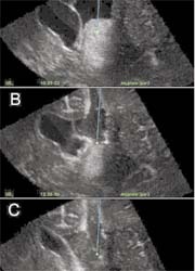
Figure 1 (small): Screen dump from the navigation system, showing corresponding image slice extracted from the MR T2 volume (top) and ultrasound volume (bottom). Note the apparent difference in tip position of the navigated instrument (cross-hair) between the images
However, conventional navigation systems, based on pre-operative images, are of limited value during surgery due to brain shift caused by the removal of cerebrospinal fluid as well as the surgical procedure. Therefore, there is a strong need for intra-operative imaging either by CT, MRI or ultrasound (US).
Intra-operative CT is not very attractive due to radiation and low sensitivity in the CT images.
Many research groups and large companies have tried to solve the brain-shift problem by using intra-operative MRI, which has proved useful for guiding certain surgical procedures and for control of tumour resection. The industry has invested in developing solutions for the operating theatre. The concept of operating inside the magnet has not been very successful due to the narrow space and need for special non-magnetic instruments. Recently, firms have made operating suites in which a patient can be transported in and out of the magnetic field. But however well designed, this transportation will interrupt an operation. Another solution is to lift small magnets in and out of the operating field (PoleStar).
Although most intra-operative MRI solutions provide good quality images, the main drawback is cost - too high for most clinics, anywhere. On top of MRI equipment operating rooms need special shielding.
Ultrasound (US) is a very interesting alternative for intra-operative imaging. In recent years US has gone through considerable technical development, mainly driven by cardiology needs. Many neurosurgeons have not recognised this development. Today, image quality from high-end US scanners is usually as good as that of pre-operative MR images (Fig 1). By some simple clinical arrangements high US image quality is also obtainable during and towards the end of an operation (Fig 2). We have used a system that integrates high-end US with neuronavigation (SonoWand). It takes under a minute to obtain a 3-D US volume, then we can immediately navigate in that volume. A new US volume can be acquired at any time during an operation. This solves our brain-shift problem, and also our need to follow the operation’s progress and the final control of the tumour resection. The system is very flexible and can be used for any operation without a time increase - rather, it reduces it, because surgeons feel more confident with the updated information.
Compared with intra-operative MR, one disadvantage of 3-D US is that it only shows a region of interest, not the whole brain. This potential problem is minimised by the navigation system, which displays corresponding image slices from both intra-operative US and pre-operative MRI (Fig 1 and 3). Another disadvantage is that neurosurgeons are less familiar with US than MRI. In our experience the learning curve is very steep.
We have used intra-operative 3-D US for several clinical applications; not just brain, spinal and skull base tumours and cavernous haemangiomas (Fig 3), but also vascular lesions (Unsgaard, G et al. Intra-operative 3D Ultrasound in Neurosurgery. Acta Neurochir. 2005 Dec 19) One important feature of intra-operative 3-D US is its ability to make 3-D angiography volumes based on power Doppler. This has been useful in tumour, AVM and aneurysm surgeries (Fig 4).
3-D US is cheap, flexible and very useful equipment for a range of neurosurgical applications. It is the workhorse of the operating theatre. We think that, because neurosurgeons are gradually discovering its usefulness, most operating theatres, worldwide, will be equipped with this type of instrument.
01.05.2006