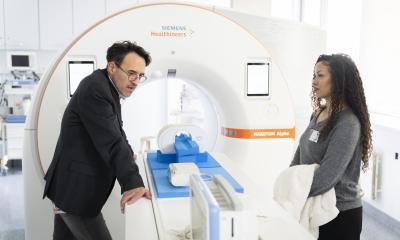
20 years of contrast enhanced MRI at Bayer Schering Pharma
Since the mid 1900s MRI has enriched clinical practice
What was initially viewed with scepticism developed into an integral and indispensable part of modern imaging diagnostics

‘At the beginning of the 1980s when Bayer Schering Pharma developed the first ever MRI contrast medium – Magnevist – some radiologists were of the opinion that conventional imaging procedures were more than enough to cover diagnostic needs. Others doubted whether contrast media were going to enhance the diagnostic significance of this new technology,’ recalls Dr Hanns-Joachim Weinmann, former head of MRI and X-ray research at Bayer Schering Pharma and a co-developer of Magnevist.
A current stock-taking appraisal belies the former scepticism: Today, in 2008, more than 20,000 MRI scanners have been installed worldwide and an annual 20 million examinations are performed – a number estimated to double over the next decade. ‘This MRI success story was only possible due to simultaneous development in three areas: scanning and coil technology, IT and contrast media,’ Professor Hans Maier, Head of the Business Unit Diagnostic Imaging, pointed out.
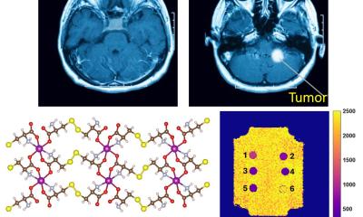
The development did not arrive from nowhere; it involved considerable work: ‘At the beginning of 1980, our first experiments were not very promising, so many colleagues ended up concentrating on the development of new contrast media for conventional X-ray procedures. It took a good year until I found a gadolinium-compound that was not toxic but still gave a clear contrast. When we could successfully diagnose a tumour in an animal brain for the first time we realised we had found the desired combination between MRI and a new type of contrast medium with optimum characteristics,’ Dr Weinmann added.
This was also the breakthrough for gadolinium-based contrast media, with the first phase of clinical trials beginning in 1983 and the initial registration granted in 1988.
The current range of products services the whole spectrum of MRI indications. It comprises the all-round Gadovist, Primovist, the first liver-specific T1 contrast medium and Vasovist, the first blood-pool marker worldwide for the visualisation of blood vessels in a dynamic and steady state.
Competition in the contrast media market today is significantly more difficult than 20 years ago, but the company also benefits from a strong therapeutic expertise alongside its diagnostics units. This is an advantageous combination, which not only facilitates modern disease management but also actively promotes it – and forms the basis for the innovations of the next 20 years.
What is MRI?
Water – the most common molecule in the human body – is the key to magnetic resonance imaging (MRI): Hydrogen atoms in the body are exposed to a strong magnetic field so that individual protons in the atomic nuclei align themselves just like the tiniest compass needles along the magnetic field. Then the protons are disturbed by radio waves. Once they return to their initial positions they send out electromagnetic signals. As these signals have specific characteristics, MRI technology can not only exactly attribute each signal to a certain position in the body, but also draw conclusions from them as to the state of the tissue.
The effect of MRI contrast media is based on paramagnetic compounds – such as the gadolinium-complex Magnevist. These produce the smallest local magnetic fields and thus change the strength of signals transmitted. In this way they enhance the contrast between the different types of body tissues, for instance between a tumour and healthy tissue.
28.10.2008