Digital mammography and tomosynthesis
`A combination of modalities is more effective in breast cancer detection´
It is hoped that a new technology, digital breast tomosynthesis or 3-D mammography, will overcome three drawbacks of traditional screening mammography: discomfort with breast compression, cancer concealed within overlapping tissue and the limited number of views.
All the major women’s imaging companies are rushing to introduce breast tomosynthesis. Boston-based Hologic appears closest to going to market. Two studies that used the Hologic technology were published recently. During the late breaking news event at the RSNA in November, Elizabeth Rafferty MD, Director of Breast Imaging at the Massachusetts General Hospital, Boston, and colleagues, reported on a reader study involving 12 radiologists who examined over 300 cases gathered from clinical sites in the USA and Europe. The researchers found that these radiologists demonstrated statistically significant better performance in reading cases with breast tomosynthesis combined with digital mammography than when using digital mammography alone. ‘Across the spectrum of age and experience, every reader performed better,’ Dr Rafferty noted.
The researchers estimated that digital breast tomosynthesis in conjunction with digital mammography can reduce the screening recall rate in a normal screening population by 40%. ‘That’s because tomosynthesis gives you the value of being able to correctly dismiss summation shadow or overlapping structures, more accurately characterise lesions and still see calcifications,’ she explained, adding that breast tomosynthesis ‘… had far more impact than we even thought it would’. The modality will ‘take away the guesswork’, she said: an important claim considering that currently-used mammography can miss some 20-30% of breast tumours. In addition, there were highly significant improvements in radiologist performance when analysing masses.
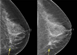
In another breast tomosynthesis study using the Hologic technology, radiologist H J Teertstra and a team at the Netherlands Cancer Institute in Antoni van Leeuwenhoek Hospital (NKI-AVL), Amsterdam, examined women to see whether the method is suitable for detecting lesions that were not seen during screening programmes that used conventional analogue mammography (see interview with Dr Teerstra, European Hospital Mammography Special, issue 4/2007). In the first 300 women examined, breast tomosynthesis detected cancer in two women that were missed with conventional digital mammography.
One major issue with 3-D imaging in general has been the belief that 3-D would bring with it an increase in radiation dose for the patient. Dr Rafferty’s team found that two rounds of digital breast tomosynthesis from different angles actually generated a lower dose of radiation than a single round of digital mammography.
As for how long it will take for breast tomosynthesis to catch on with radiologists, Dr Rafferty said: ‘That really comes down to comfort level [of the radiologist]. The fact that CAD has caught on so quickly really shows how radiologists are not confident at interpreting mammograms.’
Initially it is likely that breast tomosynthesis will be used as an adjunct to digital mammography. Dr Rafferty and colleagues, as well as Hologic, one of the manufacturers developing the new technology, believe that, if it is introduced in this way, radiologists will more easily accept the modality.
The results of these two studies strengthen Hologic’s efforts to be first to market breast tomosynthesis and enhance the company’s position that breast tomosynthesis, based on their selenium direct to digital detector, offers a superior breast tomosynthesis system. Hologic is awaiting FDA approval of digital breast tomosynthesis and believes that this equipment could become clinically available as early as mid-2008.
In addition to work on breast tomosynthesis, Hologic showcased two new Selenia digital mammography options of considerable interest at the RSNA show – Selenia S and a tungsten X-ray tube Selenia system.
Selenia S, offered at a lower price point than the popular Selenia system, is optimised for screening mammography, satellite offices or mobile environments. Selenia S can be upgraded to include all diagnostic tools found on Selenia.
While radiation exposures in digital mammography are already very low relative to the patient benefit from early breast cancer detection, Hologic offers a tungsten X-ray tube with rhodium and silver filters Selenia system. The use of a tungsten X-ray tube enables dose reductions of around 30%, while maintaining the excellent image quality already achieved with Selenia, Hologic reports. ‘The new system also offers superior performance for some of the advanced applications under development, such as digital breast tomosynthesis, iodinated contrast, and dual energy breast imaging.’
31.12.2007